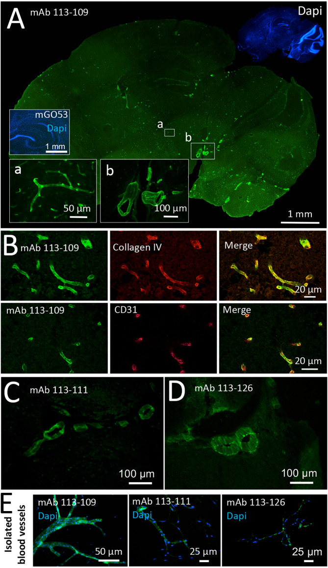
Figure 1.
Recombinant monoclonal antibodies from a GABAAR encephalitis patient bind to brain blood vessels. Sections from unfixed and unpermeabilized adult mouse brains were incubated with 5 μg/ml of the respective monoclonal antibodies obtained from a patient diagnosed with GABAAR encephalitis. Visualization of tissue binding was performed using a FITC-coupled anti-human IgG secondary antibody. (A) Sagittal brain section incubated with human monoclonal antibody (mAb) 113-109. Prominent staining of blood vessels of all diameters in all brain regions was obtained (see insets a and b). Incubation with control mAb mGO53 yielded no tissue staining (inset). (B) Double stainings against Collagen IV (upper panel) and CD31 (lower panel) confirm immunoreactivity of mAb 113-109 to blood vessels. (C) Sagittal brain section incubated with mAb 113-111. Prominent staining of large blood vessels was obtained in all brain regions (the area between the cerebellum and midbrain is shown). (D) Sagittal brain section incubated with mAb 113-126. Again, prominent staining of large blood vessels in all brain regions was obtained (the area between the cerebellum and midbrain is shown). (E) Stainings of isolated brain blood vessels. Blood vessels were obtained from homogenized mouse brains by combinatory centrifugation and filtering steps. A 30-100 μm filter size fraction incubation with all three antibodies resulted in clear staining of the vessels within this fraction. Note that the diameter of mounted blood vessels is subject to shrinking artifacts during the staining procedure.

