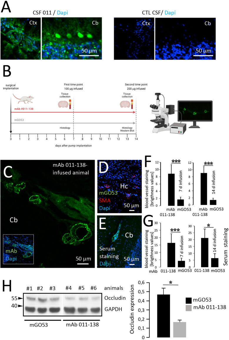
Figure 4.
Intrathecal application of antibody 011-138 leads to in vivo blood vessel binding and Occludin downregulation. (A) Sections from unfixed adult mouse brains were incubated with CSF of patient 011 and an age-matched control patient at a dilution of 1:2. Incubation with 011 CSF resulted in IgG staining of blood vessels and cerebellar Purkinje cells. Incubation with control CSF yielded no staining. (B) Adult mice were either administered a dose of 100 μg of human monoclonal antibody (mAb) 011-138 into the right lateral ventricle for 7 days or 200 μg for 14 days. Animals were sacrificed, brains were removed, and immediately frozen for immunohistochemistry. In addition, blood was collected to obtain serum. (C) Representative sagittal brain section from an animal treated for 14 days with mAb 011-138 was incubated with FITC-coupled anti-human IgG. Clear staining of large to mid-size blood vessels in all brain regions was visible (shown for the cerebellum, Cb). (D) Sagittal brain section from one animal treated with control antibody mGO53 for 14 days. Incubation with a secondary antibody revealed no staining Hc = hippocampus. (E) Sagittal unfixed brain section from an untreated adult mouse was incubated with serum (dilution 1:200) from an animal that had received mAb 011-138 for 14 days. Visualization with secondary antibody revealed staining of larger to mid-size blood vessels by the mouse serum. (F) Quantification of blood vessel IgG immunoreactivity. Data are given as means ± SEM from three animals per condition. Per condition, between 38 and 43 blood vessel sections were analyzed. ***p ≤ 0.001. (G) Quantification of serum blood vessel IgG immunoreactivity on naive brain sections. Data are given as means ± SEM from three animals per condition. Per condition, between 22 and 45 blood vessel sections were analyzed. *p ≤ 0.05, ***p ≤ 0.001. Brightness levels of mGO53 staining were within the background range. (H) Downregulation of Occludin by mAb 011-138. In brains of mice treated with mAb 011-138 for 14 days protein expression was strongly reduced by 65% compared to animals that had received mGO53. Data are given as means ± SEM adjusted to loading from seven animals per condition from two independent experiments. *p ≤ 0.05. Three animals per condition from one experiment are exemplarily shown.

