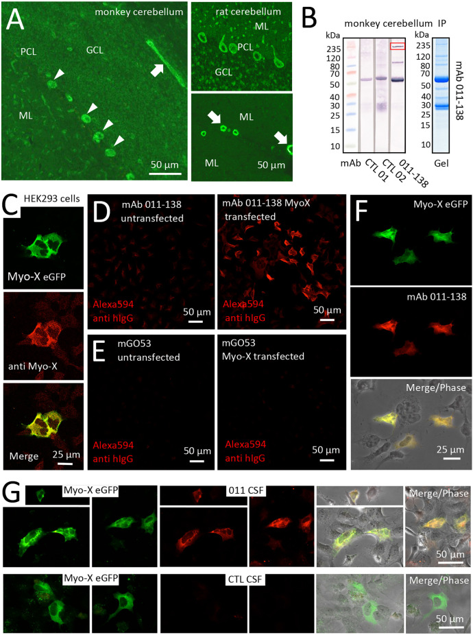
Figure 5.
Antibody 011-138 targets Myosin-X in transfected HEK cells. (A) Monkey and rat brain biochip cryosections (EUROIMMUN AG) were incubated with human monoclonal antibody (mAb) 011-138 (1:100) and exhibited pronounced staining of blood vessels (left panel and lower right panel arrows) and Purkinje cells (left panel arrowheads and upper right panel). (B) Immunoprecipitation analysis using monkey brain lysates. Lysates were incubated with either mAb 011-138 or two other non-vessel reactive human monoclonal antibodies for control. Dynabeads were used to precipitate antibodies together with bound antigens. Elute fractions were again incubated with the precipitating antibody followed by Western blotting. The marked band at 55 kDa in all three lanes presumably resulted from detection of the precipitating heavy chain by the detecting secondary anti-human IgG conjugate. In addition, mAb 011-138 showed two distinct bands at 80 and 240 kDa (boxed), respectively. The corresponding Coomassie gel is shown for mAb 011-138. (C) HEK 293 cells were transfected with an eGFP construct of human Myosin-X (Myo-X) plasmid DNA for 24 h. Expression of Myo-X was verified in fixed cells using a commercial monoclonal anti-Myo-X antibody showing a very high degree of signal overlap (confocal imaging). (D) Incubation of Myo-X-transfected cells with 5 μg/ml of 011-138 antibody showed binding to fixed transfected cells that were absent in untransfected cells. A secondary anti-human IgG antibody coupled to Alexa594 was used for detection. (E) For negative control untransfected and Myo-X-transfected cells were incubated with 5 μg/ml of mGo53 antibody. No staining was observed under either condition. (F) In transfected cells incubated with 011-138 antibody the human IgG signal showed a very high degree of overlap with eGFP-Myo-X signal. (G) HEK 293 cells were transfected with an eGFP construct of human Myo-X plasmid DNA for 24 h. Incubation of Myo-X-transfected cells with patient-CSF 011 showed binding that was absent after incubation with control-CSF. The right panel shows a high degree of overlap of patient-CSF 011 signal with eGFP-Myo-X signal.

