Figure 6.
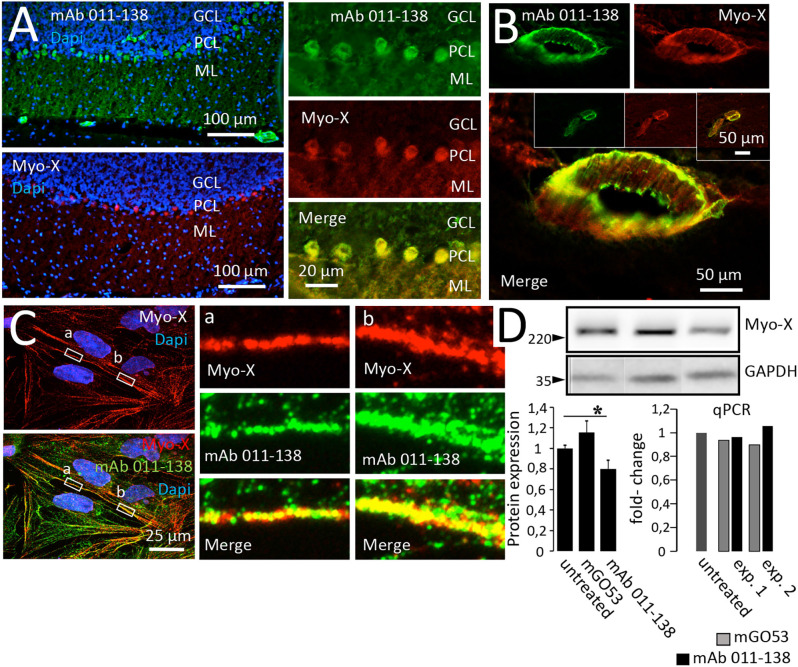
Antibody 011-138 colocalizes with commercial anti-Myosin-X antibodies in brain sections and hCMEC/D3 cells and downregulates Myosin-X. (A) Unfixed and unpermeabilized sections from adult mouse brains were incubated with 5 μg/ml of human monoclonal antibody (mAb) 011-138 and a commercial monoclonal mouse antibody directed against unconventional Myosin-X (Myo-X). Shown is the 3-layer cerebellar cortex consisting of the granule cell layer (GCL), Purkinje cell layer (PCL), and outermost molecular layer (ML). Incubation with mAb 011-138 resulted in staining of the Purkinje cell somata and larger blood vessels (upper panel). Staining with commercial anti-Myo-X antibody showed a comparable pattern (lower panel, no larger vessels present). Double staining revealed a high degree of signal overlap between patient 011-138 and commercial MyoX antibodies in Purkinje cells. (B) Likewise, double staining revealed a high degree of signal overlap between patient 011-138 and commercial Myo-X antibodies in mid-size (insets) to larger blood vessels. (C) Double incubation of fixed hCMEC/D3 cells with antibody 011-138 and commercial anti-Myosin-X IgG. Both stainings yielded a similar staining pattern with partially overlapping signals (see insets). (D) Incubation of hCMEC/D3 cells with mAb 011-38 results in the downregulation of Myo-X. Following incubation of hCMEC/D3 cells with 5 μg/ml of mAb 011-138 or mGO53 as control for 48 h cells were homogenized and subjected to Western blotting and quantitative PCR to check for protein expression and gene regulation. Incubation with mAb 011-138 decreased protein expression of Myo-X by 20% (left chart), gene regulation was unaltered (right chart). Data are given as normalized means ± SEM adjusted to loading from two individual experiments analyzed in duplicates (Western blot) and as two individual experiments (qPCR). *p ≤ 0.05.

