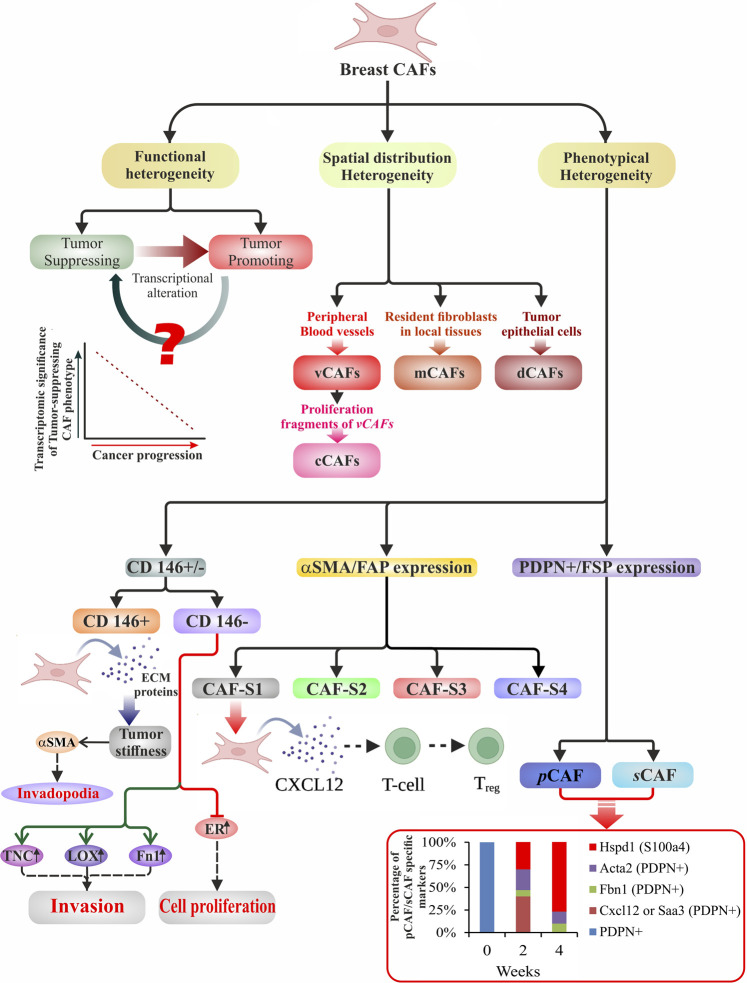
FIGURE 1.
Schematic illustration of heterogeneity in breast CAFs. Broadly, breast CAFs are distinguished under three subclasses based on their heterogeneous function, spatial distribution, and cell surface phenotype. They have been divided into tumor-promoting and tumor-suppressing cell types due to their stage-specific distinct approach toward tumor cells. With the progression of cancer, transcriptional alteration of the tumor-suppressing CAF phenotypes results in the generation of tumor-promoting breast CAFs. Manipulation of the event may prove to be a potent therapeutic tool. Bartoschek et al. (2018) used single-cell RNA data to classify the CAFs according to their spatial distribution. Vascular development, ECM-enriched signaling, expression of proliferative genes, and variously expressed differentiation genes shaped their classification into vCAFs, mCAFs, cCAFs, and dCAFs. Phenotypically breast CAFs could be sub-classified depending upon the i) presence or absence of CD146 (Brechbuhl et al., 2020), ii) expression of αSMA/FAP (Costa et al., 2018), and iii) expression of PDPN/FSP (Friedman et al., 2020). This diagram displays the several subtypes with phenotypical distinctions that are accountable for classified functions at various phases of carcinogenesis. The bottom right graph illustrates the percentage of pCAF/sCAF specific markers over a period of 4 weeks in a breast cancer model.

