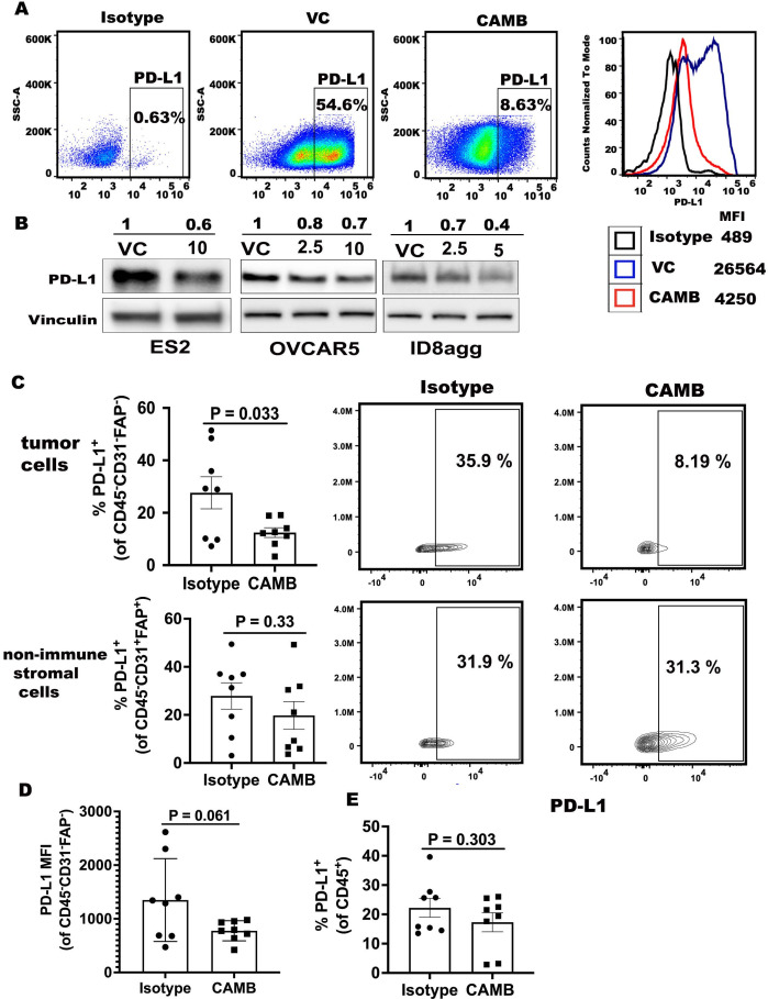
Figure 1.
Chlorambucil reduces tumor PDL1 in vitro and in vivo. (A) Flow cytometry for PDL1 percentage and mean fluorescence intensity (MFI) in VC (vehicle control) and chlorambucil treatment (10 µM, 48 hours) in OVCAR5 cells in vitro. (B) Western blots for PDL1 expression in human and mouse ovarian cancer cells treated in vitro with (+) or without (−) chlorambucil for 48 hours. Vinculin, loading control. Values are μM chlorambucil. (C) Flow cytometry analyses of WT mice challenged with ID8agg cells, treated with chlorambucil (CAMB, 1 mg/kg as described in methods) and sacrificed 4 weeks post tumor challenge. Summary of PDL1 percentage in the tumor CD45-CD31-FAP- (top) or non-immune stromal cell CD45-CD31+FAP+ (bottom) gates. PDL1 percentage in specified populations as indicated (right). (D) Summary graph of PDL1 MFI in CD45-CD31-FAP- tumor cells. (E) Summary of PDL1 percentage in CD45+ immune cells. P value determined by unpaired t-test. VC, vehicle control.

