. 2012 Mar;129(3):e748–e755. doi: 10.1542/peds.2011-2067

FIGURE 2.

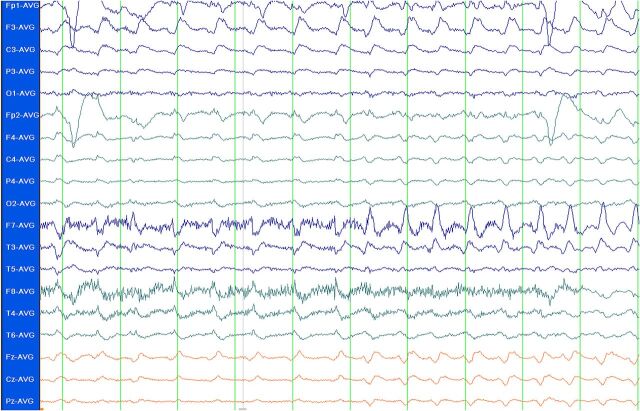
FIGURE 2. EEG showing SE. Standard 10-20 system EEG in average referential montage display, demonstrating rhythmic sharps and δ frequency ictal activity maximal in the left frontal region. Patient was a 2-year-old girl with cardiopulmonary arrest secondary to pulmonary hemorrhage. She became less responsive several days after her arrest. An MRI demonstrated multifocal cortical signal abnormalities consistent with cerebral edema. This EEG was obtained, and a diagnosis of NCSE was made. She was eventually stabilized but on follow-up had significant neurologic impairment.

EEG showing SE. Standard 10-20 system EEG in average referential montage display, demonstrating rhythmic sharps and δ frequency ictal activity maximal in the left frontal region. Patient was a 2-year-old girl with cardiopulmonary arrest secondary to pulmonary hemorrhage. She became less responsive several days after her arrest. An MRI demonstrated multifocal cortical signal abnormalities consistent with cerebral edema. This EEG was obtained, and a diagnosis of NCSE was made. She was eventually stabilized but on follow-up had significant neurologic impairment.