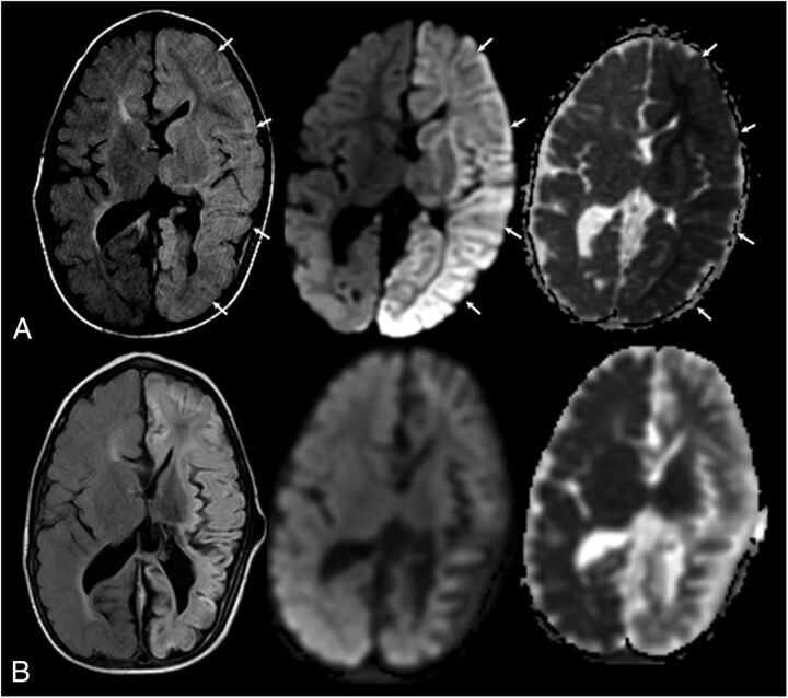
FIGURE 3.
A 2-year-old boy with new-onset seizures and sepsis. History of previous white matter injury of prematurity and intraventricular hemorrhage. Shunt dependent hydrocephalus. L-R: Axial fluid attenuated inversion recovery image, axial diffusion-weighted image, and apparent diffusion coefficient map. A, At acute clinical presentation. Note the extensive edema and gyral swelling throughout the left hemisphere on the fluid attenuated inversion recovery sequence (arrows). Increased signal is also noted involving the left thalami and basal ganglia. Trace diffusion-weighted image map demonstrates extensive increased signal throughout the left hemisphere. Apparent diffusion coefficient map demonstrates left hemispheric diffusion restriction involving both the cortex and white matter. Follow-up imaging (1 month later) demonstrates diffuse left hemispheric volume loss and evolution of diffusion changes. Clinically, he had acute flaccid right hemiplegia. At 6-month follow-up, he was improved but continued to have persistent spastic right hemiparesis. Language developmental milestones were normal.

