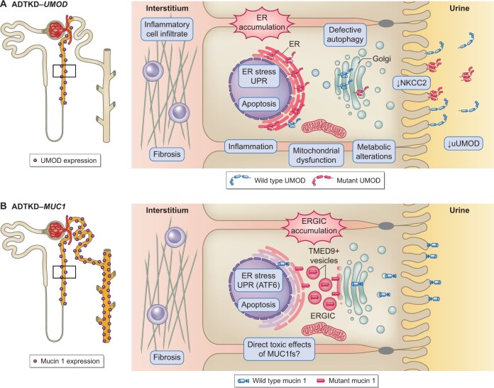
FIGURE 1.
Pathophysiology of ADTKD-UMOD and ADTKD-MUC1. (A) Left panel: UMOD expression is restricted to the thick ascending limb and the early DCT. Right panel: Cellular pathways activated by the expression of mutant UMOD (in cellular and murine studies as well as patient biopsies) and its toxic accumulation in the ER. (B) Left panel: Mucin 1 expression along the entire distal tubule. Right panel: Cellular pathways activated by the expression of mutant mucin 1 (in cellular and murine studies as well as patient biopsies) and its toxic accumulation in the ERGIC.

