Figure 3.
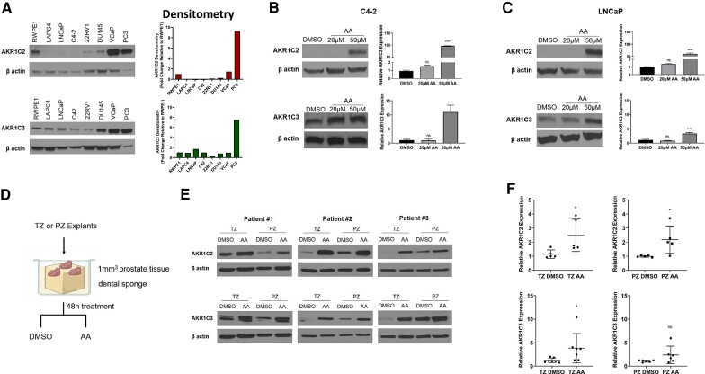
Arachidonic acid (AA) induces expression of AKR1C2 and AKR1C3. (A) Endogenous protein expression and quantification of AKR1C2 (top panel) and AKR1C3 (bottom panel). (B, C) Protein and transcript expression of AKR1C2 and AKR1C3 in C4-2 and LNCaP cells treated with DMSO (control) or AA (20 μM and 50 μM) for 24 hours. (D) Schematic of ex vivo culture of prostate tissue explants from the transitional zone (TZ) and peripheral zone (PZ) treated with DMSO (control) or AA (50 μM). (E) Protein and transcript (F) expression of TZ and PZ tissue explants treated with DMSO or AA for 48 hours. Western blots were quantified with ImageJ and are representative of at least 5 biological replicates. *P < 0.05, **P < 0.01, ***P < 0.001, ****P < 0.0001, ns = not significant.

