
Figure 1. Multiplexed reporter system to monitor MAPK activity and dynamics in single cells.
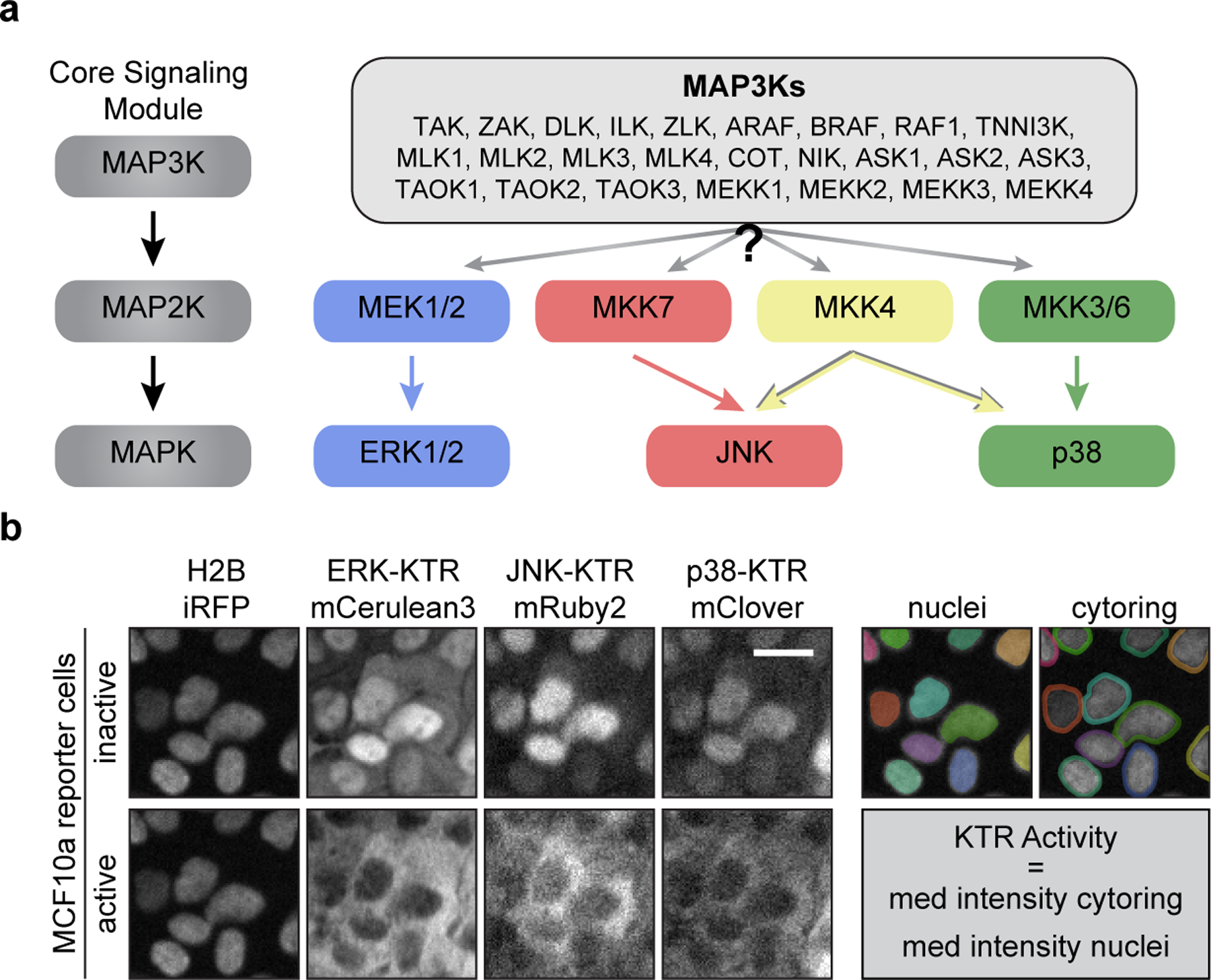
a. Schematic of the MAPK core signaling module. MAP3Ks phosphorylate and activate MAP2Ks which in turn activate MAPKs (left). Schematic representing the mammalian MAPK network, where one of the listed MAP3Ks may activate one or more MAP2K which in turn activate MAPKs (right). b. Representative images of MCF10a cells expressing H2B-iRFP, ERK-KTR-mCerulean3, JNK-KTR-mRuby2, and p38-KTR-mClover before and after treatment with sorbitol (100 μM) (left). Diagram of ‘nuclei’ and ‘cytoring’ segmentation where each individual cell is marked with another color (right). Scale bar = 20 μm.