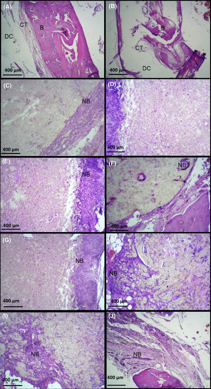
Fig. 8.

Hematoxylin eosin staining at 6 (first column, A, C, E, G, K) and 12 (second column, B, D, F, H, L) weeks after implantation (scale bar: 400 µm). The results showed that new bone like tissue (NB) expanded to the scaffolds at defects filled by PTFE/PVA (C, D), PTFE/PVA/GO (E, F), PTFE/PVA/Cell (G, H), and PTFE/PVA/GO/Cell (K, L) as compare to control group (A, B). Host bone (B), Defect cavity (DC), Connective tissue (CT), Osteocyte (OSC), New bon like form tissue (NB).
