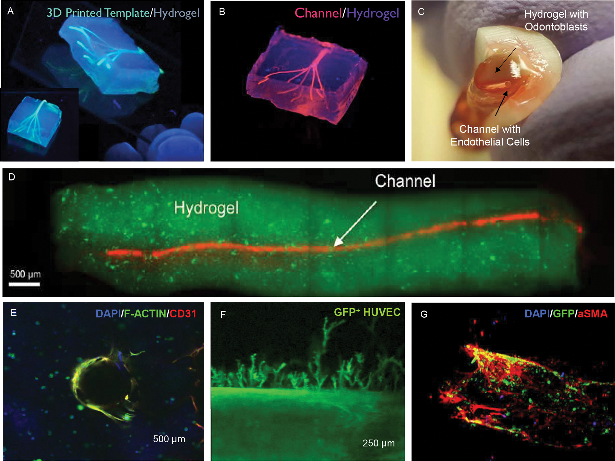
Figure 6. 3D printing and microengineering of the dental pulp vasculature.

(A) Extrusion 3D printing of sacrificial template fibers embedded in cell-laden hydrogels followed by (B) template fiber removal resulting in the formation of bifurcating channels in the range of 100–1000 μm. Reproduced from (53) (C-D) A similar strategy of sacrificial templating was adapted to microengineer hollow conduits embedded in odontoblast-laden photocrosslinkable hydrogels loaded into the root canal space of full-length human teeth. Reproduced from (38). (E) These hydrogel conduits can be loaded with endothelial cells to form a monolayer that can (F) lead to endothelial sprouting into the hydrogel matrix. (G) When endothelial cells (HUVECs) are co-cultured with bone marrow mesenchymal stromal cells in the right conditions, a pericyte-supported vascular channel can also be formed.
