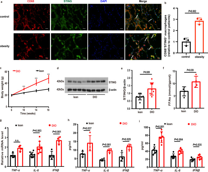
Fig. 1. The levels of STING and proinflammatory cytokines were increased in obesity.
a Representative lung immunofluorescence images of STING+/CD68+ macrophages from patients with obesity and controls. Scale bars represent 25 μm. b Quantification analysis of the numbers of STING+/CD68+ macrophages in patients with obesity and controls (n = 3 each group). c Body weight of regular chow (n = 14) and high-fat diet (n = 15) fed mice. d, e Representative western blots of STING expressions (d), and (e) group data of STING fold change in equal amounts of protein extracts of lung tissues from lean (n = 8) and DIO (n = 9) mice. f The levels of free fatty acids (FFAs) in lung tissues were significantly increased in DIO compared to lean mice (n = 4 each group). g qPCR analysis showed the mRNA levels of TNF-α (n = 4), IL-6 (n = 8) and IFNβ (n = 4) in DIO and control mouse lung tissues. h ELISA analysis showed that the protein levels of proinflammatory cytokines in lung homogenates were increased in DIO mice (n = 4 each group). i ELISA analysis showed that the levels of proinflammatory cytokines in BALF were increased in DIO mice (n = 6 each group). Data represent mean ± SD. Statistical significance was determined using two-tailed Student’s t test or Mann-Whitney test.

