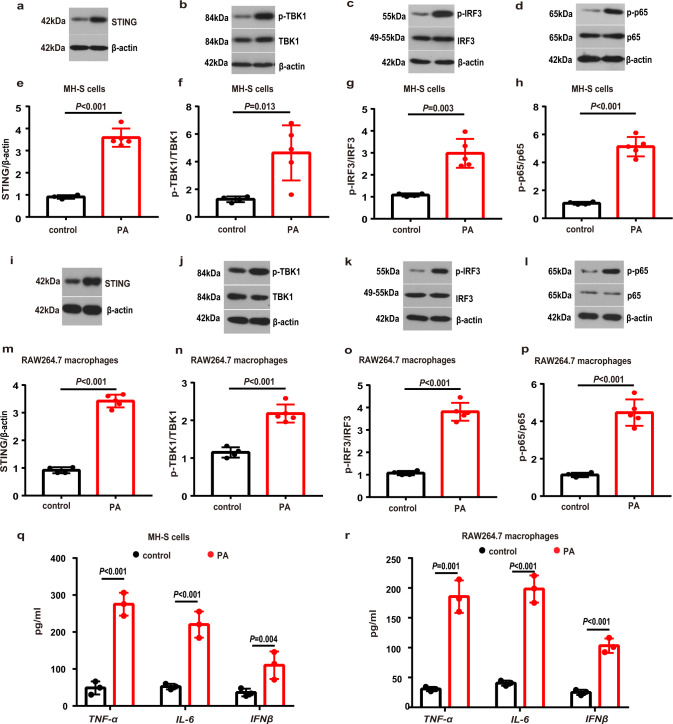
Fig. 4. PA activated STING-TBK1-IRF3/NF-κB pathway and induced inflammation in MH-S and RAW264.7 macrophages.
MH-S cells were treated with PA (0.6 mM) for 24 h. a–d Representative western blots of the STING (a), p-TBK1 (b), TBK1 (b), p-IRF3 (c), IRF3 (c), p-p65 (d) and p65 (d) in PA-treated MH-S cells. e–h Group data of STING (e), p-TBK1/TBK1 (f), p-IRF3/IRF3 (g) and p-p65/p65 (h) fold change in equal amounts of protein extracts of control (n = 4 biological repeats) and PA-treated (n = 5 biological repeats) MH-S cells. RAW264.7 macrophages were treated with PA (0.6 mM) for 24 h. i–l Representative western blots showed the STING (i), p-TBK1 (j), TBK1 (j), p-IRF3 (k), IRF3 (k), p-p65 (l) and p65 (l) in PA-treated RAW264.7 macrophages. m–p Group data of STING (m), p-TBK1/TBK1 (n), p-IRF3/IRF3 (o) and p-p65/p65 (p) fold change in equal amounts of protein extracts of control (n = 4 biological repeats) and PA-treated (n = 5 biological repeats) RAW264.7 macrophages. q–r ELISA analysis showed that the protein levels of TNF-α, IL-6 and IFNβ in MH-S cells (q) and RAW264.7 macrophages (r) were significantly increased after PA treatment. n = 3 biologically independent experiments. Data represent mean ± SD. Statistical significance was determined using two-tailed Student’s t test.

