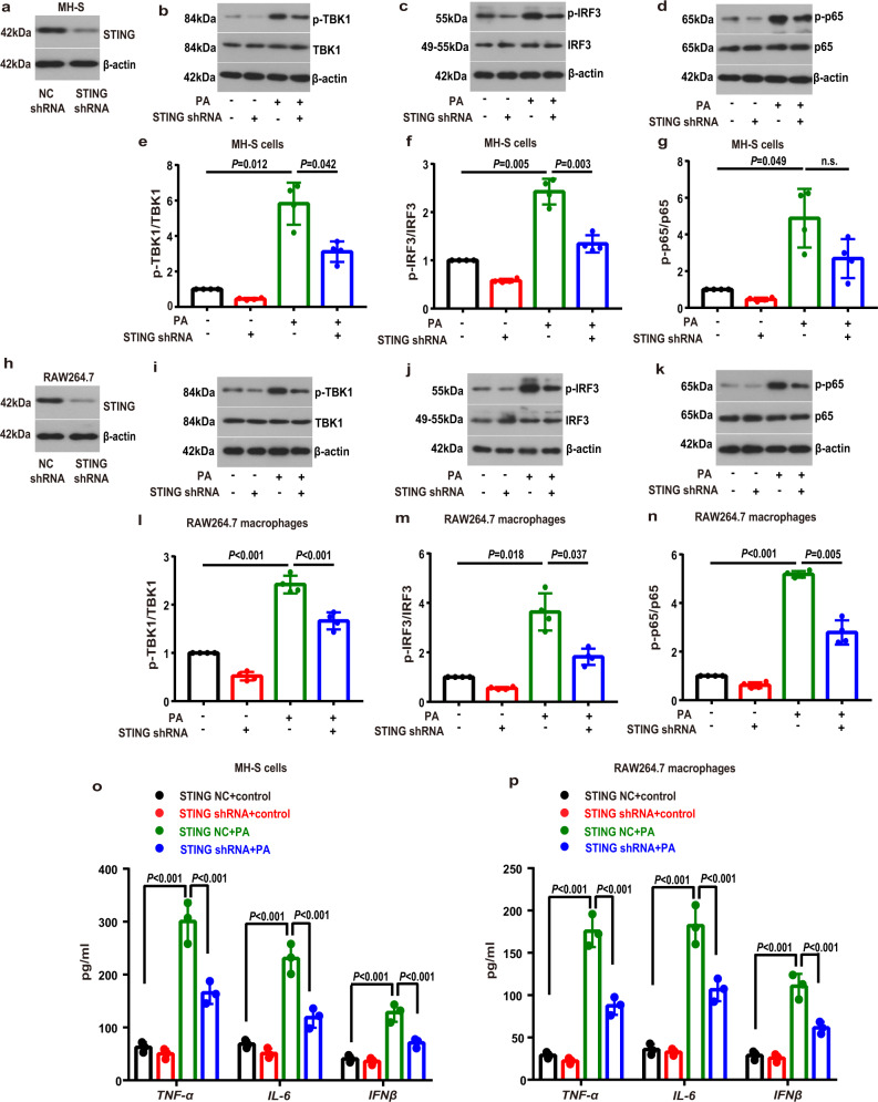
Fig. 5. STING knockdown attenuated the PA-induced inflammatory activation in MH-S and RAW264.7 macrophages.
The expression of STING was silenced by using specific shRNAs in MH-S (a) and RAW264.7 macrophages (h). Negative control (NC) shRNA was used in parallel for the control groups. b–g MH-S cells were transfected with STING shRNA or NC shRNA and then treated with PA (0.6 mM) for 24 h. Representative western blots of the p-TBK1 (b), TBK1 (b), p-IRF3 (c), IRF3 (c), p-p65 (d) and p-65 (d), and group data of p-TBK1/TBK1 (e), p-IRF3/IRF3 (f) and p-p65/p65 (g) fold change in equal amounts of protein extracts of STING shRNA or NC shRNA MH-S cells with or without PA. n = 4 biologically independent experiments in each group. i–n RAW264.7 macrophages were transfected with STING shRNA or NC shRNA and then treated with PA (0.6 mM) for 24 h. Representative western blots of the p-TBK1 (i), TBK1 (i), p-IRF3 (j), IRF3 (j), p-p65 (k) and p-65 (k), and group data of p-TBK1/TBK1 (l), p-IRF3/IRF3 (m) and p-p65/p65 (n) fold change in equal amounts of protein extracts of STING shRNA or NC shRNA RAW264.7 macrophages with or without PA. n = 4 biologically independent experiments in each group. o, p ELISA analysis showed that STING shRNA attenuated the PA-induced increase in levels of proinflammatory cytokines, including TNF-α, IL-6 and IFNβ in PA-treated MH-S cells (o) and RAW264.7 macrophages (p). n = 3 biologically independent experiments. Data represent mean ± SD. n.s. not significant. Statistical significance was determined by one-way ANOVA followed by post-hoc LSD-t or Games-Howell method.

