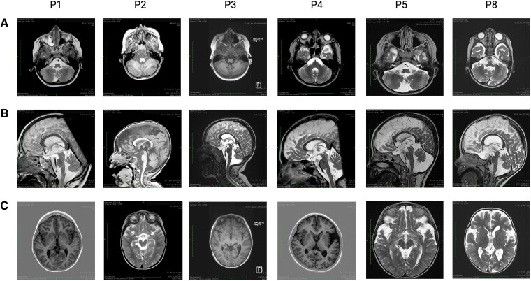
Figure 1.
Homozygosity for the p.C112Wfs*11 SOD1 variant (C112XHom) is associated with distinct cranial MRI alterations. Images display coronal and sagittal MRI scans from C112XHom patients 1, 2, 3, 4, 5, and 8. (A) Atrophy of the cerebellar vermis was seen to varying degrees in all analyzed patients. In those individuals who underwent serial imaging studies, the findings progressed over time (Table 2). (B) Brain stem atrophy was also observed in all 6 patients. (C) In 5/6 patients, some fronto-temporal atrophy was found, which did not progress visibly with age (range of observation period from 9 months to 12 years) in the three patients with serial studies.

