Figure 6.
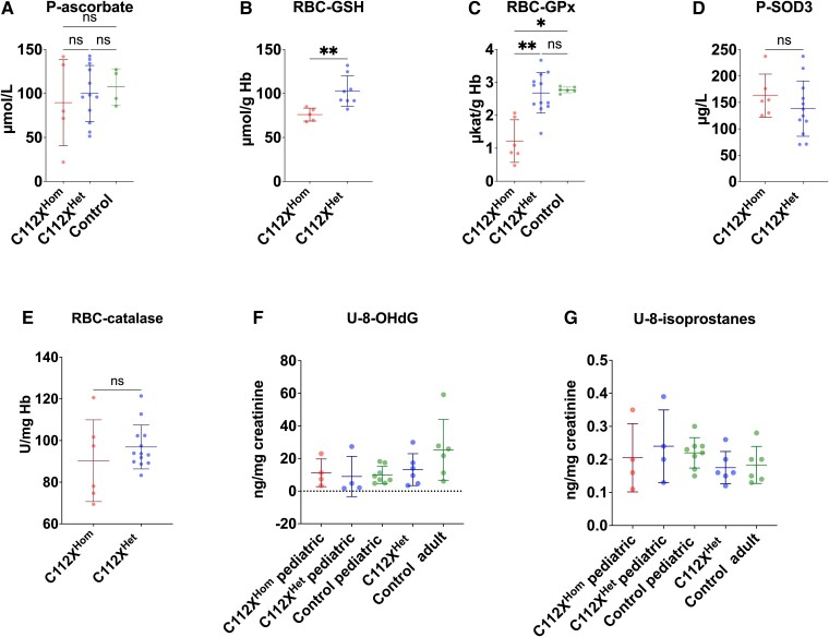
SOD1 deficiency leads to altered glutathione metabolism, while commonly used markers for oxidative stress remain unaltered. (A) Despite high reactivity with superoxide, plasma ascorbate (P-ascorbate) levels were not significantly different between homozygous (C112XHom) and heterozygous individuals (C112XHet) or controls. C112XHomn = 5, C112XHetn = 12, control n = 4. (B) In contrast, whole blood reduced glutathione (RBC-GSH) levels were significantly reduced in homozygous individuals (76 ± 7.4 µmol/g Hb, P = 0.002) when compared to heterozygous carriers (102.8 ± 17.4 µmol/g Hb). C112XHomn = 5, C112XHetn = 8. (C) Levels of the H2O2-metabolizing GSH-utilizing glutathione peroxidase-1 (RBC-GPx) were significantly reduced in homozygous individuals (1.2 ± 0.6 µkat/g Hb) when compared to both heterozygous carriers (2.7 ± 0.6 µkat/g Hb, P = 0.003) and controls (2.8 ± 0.1 µkat/g Hb, P = 0.01). C112XHomn = 6, C112XHetn = 12, control n = 6. (D) The levels of extracellular SOD3 were not significantly different between homozygous (162.8 ± 41 µg/L) or heterozygous individuals (138.4 ± 52.5 µg/L) (P = 0.26, reference range 142 ± 43 µg/L). C112XHomn = 6, C112XHetn = 12. (E) No significant difference In the activity of the H2O2-metabolizing RBC-catalase was identified between homozygous patients and heterozygous carriers (90.3 ± 19.6 U/mg Hb and 96.9 ± 10.5 U/mg Hb, respectively, P = 0.43). C112XHomn = 6, C112XHetn = 12. (F, G) Despite distinct alterations in redox metabolism, no overt differences were seen in the urinary excretion of 8-hydroxydeoxyguanosine (U-8-OHdG), a marker of oxidative damage to DNA, and urinary (U-)8-isoprostanes, a marker of lipid peroxidation. C112XHomn = 4, C112XHet paediatric n = 4, C112XHet adult n = 6, control paediatric n = 8, control adult n = 6. Data were compared using the Mann–Whitney U test (A-E) or Kruskal–Wallis test (F, G). *P < 0.05, **P < 0.01, ns: not significant.

