Figure 2.
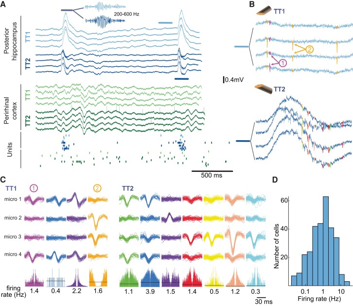
FR and spikes in local and global circuits. (A) LFP recordings (unfiltered broadband signal) on two hybrid electrodes in Subject 2. Two IEDs are visible in the posterior hippocampus, one in the perirhinal cortex. IEDs were associated with FRs, as shown in the inset (200–600 Hz filtered LFP trace). Bottom, raster plot of neuronal activity. Each row corresponds to one unit and each dot to one action potential. There is an increase in firing in most units during the IED and FR recording on TT1 and TT2 located in the posterior hippocampus (blue and purple dots). (B) Action potentials recorded on the posterior hippocampus on TT1 and TT2 immediately before the highlighted FR in A (TT1) and during the FR (TT2). Note the FR in the broadband on the TT2 trace. (C) Sample waveforms of action potentials recorded on TT1 and TT2 in the posterior hippocampus, sorted into putative single units. Bottom, auto-correlogram (± 30 ms) and mean firing rate of each single unit. (D) Distribution of single unit firing rates (log scale).

