
FIGURE 5.
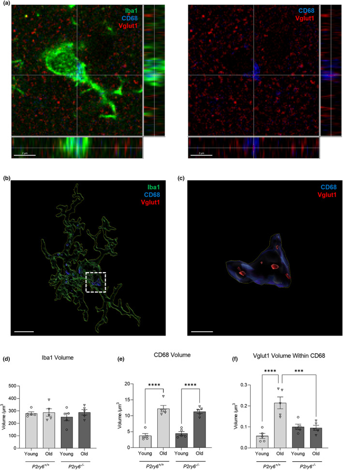
P2Y6R deficiency prevents age‐associated synaptic phagocytosis. (a) Representative confocal microscopy image of mice stained for Iba1 (green, microglial marker), CB68 (blue, lysosomal marker), and Vglut1 (red, synaptic marker) in the somatosensory cortex. Scale bar = 2 μm. (b) Representative surface‐rendered microglia (from a). Scale bar = 3 μm. (c) Enlarged inset of Vglut1 colocalization within CD68, denoted by the white dotted line (from b). Scale bar = 0.5 μm. Microglial volume (d), CD68 volume within microglia (e), and Vglut1 colocalization within CD68 (f) across young (4 months) and old (17 months) wild‐type and knockout mice. Each point represents one animal comprised of 13–15 microglia analyzed across three equidistant sections. Statistical comparisons were made via a two‐way ANOVA with Bonferroni's post hoc comparison test. Error bars represent ±SEM, ***p < 0.001, ****p < 0.0001