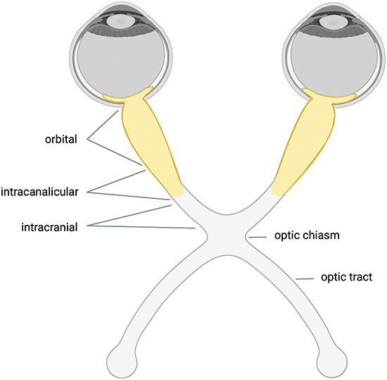
FIG. 2.

Optic nerve lesions in MOGAD. In MOGAD, bilateral ON is observed in up to 45% of patients and optic disc edema is common. MRI shows enhancement of the optic nerve and perineural abnormalities including optic nerve sheath enhancements in half of the patients. Furthermore, lesions are usually longitudinal extensive and affect predominantly the prechiasmic optic nerve (highlighted in yellow). Involvement of the optic chiasm and the optic tract is only observed in 12% and 2% of patients, respectively. Data from (Refs. 141,143). Created with BioRender.com. MOGAD indicates myelin oligodendrocyte glycoprotein-associated disease; ON, optic neuritis
