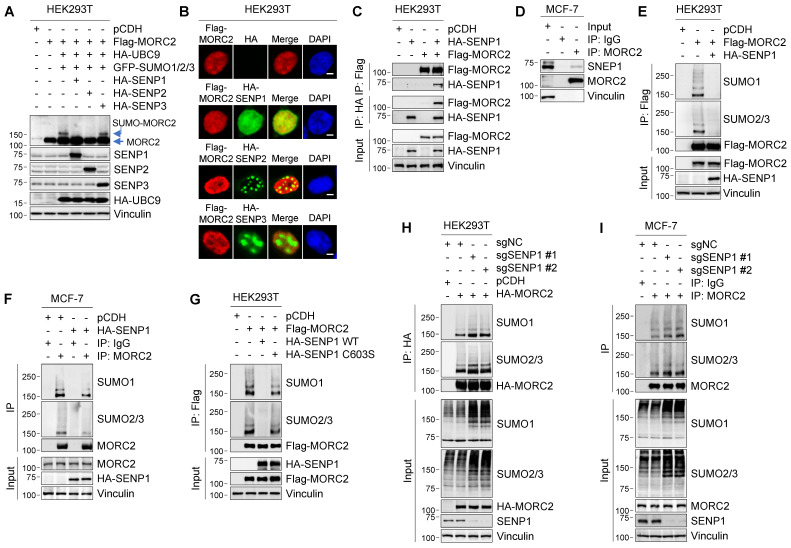
Figure 4.
SENP1 serves as a deSUMOylase for MORC2. (A) HEK293T cells were transfected with the indicated expression vectors. Cellular lysates were analyzed by immunoblotting. (B) Co-localization of MORC2 with HA-SENP1, HA-SENP2 or HA-SENP3 was analyzed by immunofluorescent staining with the indicated antibodies. Nuclei were counterstained with DAPI. Scale bar: 2.5μm. (C) HEK293T cells were transfected with Flag-MORC2 and HA-SENP1 alone or in combination. Cellular lysates were subjected to IP assays with anti-Flag or anti-HA beads, followed by immunoblotting analysis with the indicated antibodies. (D) Lysates from MCF-7 cells were processed for IP assays using an anti-MORC2 antibody, and immunoblotted with indicated antibodies. (E-F) HEK293T (E) and MCF-7 (F) cells were transfected with the indicated expression vectors. The sequential IP and immunoblotting analyses were conducted with the indicated antibodies. (G) HEK293T cells were transfected with Flag-MORC2 alone or in combination with WT or C603S HA-SENP1. The sequential IP and immunoblotting analyses were conducted with the indicated antibodies. (H-I) Endogenous SENP1 was knocked out using the CRISPR/Cas9 system in HEK293T cells expressing HA-MORC2 (H) or in MCF-7 cells (I). The sequential IP and immunoblotting analyses were conducted with the indicated antibodies.

