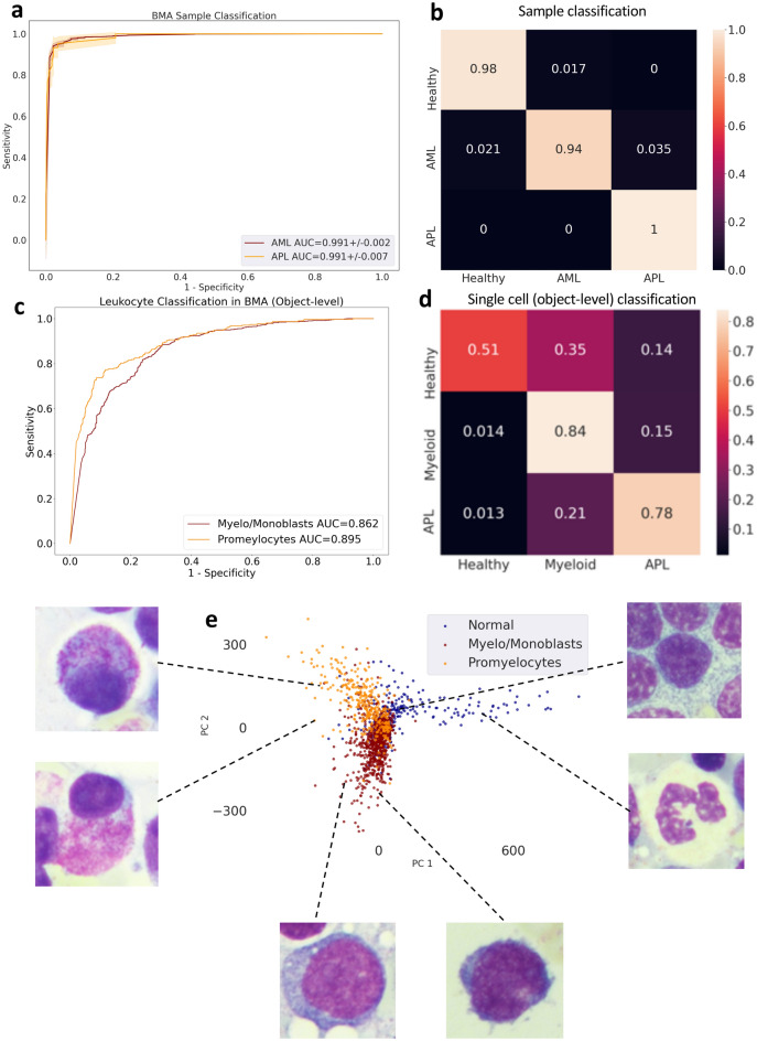
Figure 5.
Acute myeloid leukemia detection in bone marrow aspirates. (a) Receiver operating characteristic (ROC) curve for sample-level classification on the hold out validation set. (b) Confusion matrix for sample classification on the validation set. (c) ROC curve for cell-level classification on the cell image test set. (d) Confusion matrix for cell classification (promyelocytes vs other) on the cell image test set. (e) PCA visualization of the convolutional representations learned by MILLIE of the individual cells in the test set.

