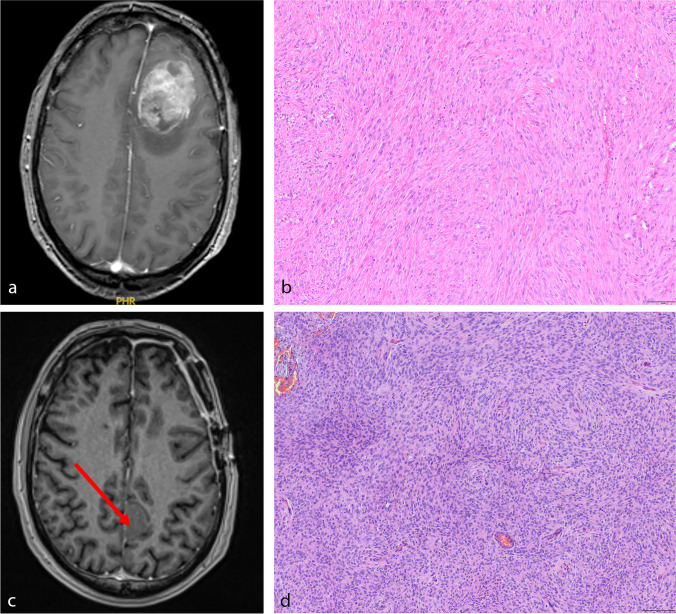
Fig. 2.
Illustrative sample of a patient developing metachronous spatially distinct meningiomas. After resection of a left frontal parasagittal meningioma (a, axial T1-weighted, contrast-enhanced imaging), microscopic analyses revealed transitional meningioma, WHO grade 1, b). Four years later, the patient developed a left parietal, parasagittal lesion (axial T1-weighted, contrast-enhanced imaging, c, arrow), diagnosed as atypical meningioma (d, hematoxylin and eosin staining, each)

