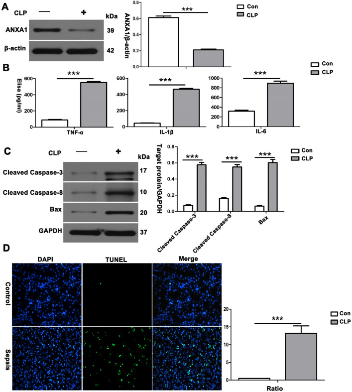
Fig. 1.
Reduction in ANXA1 produced a significant increase in inflammatory cytokines, proapoptotic proteins and apoptosis in kidney tissue in mice. A Representative western blots and quantification results of ANXA1 after CLP for 24 h. β-Actin was used as a loading control for the total protein. B TNF-α, IL-1β and IL-6 levels in the serum of mice were determined by ELISA after CLP for 24 h. C Representative western blots and quantification results of cleaved caspase-3, cleaved caspase-8 and Bax after CLP for 24 h. GAPDH was used as a loading control for the total protein. D Renal tissue apoptosis was determined by TUNEL after CLP for 24 h. Note: Con: control group, CLP: CLP group. The data are represented as the mean ± SEM. The data were independently replicated in triplicate. ***P < 0.001

