Fig. 6.
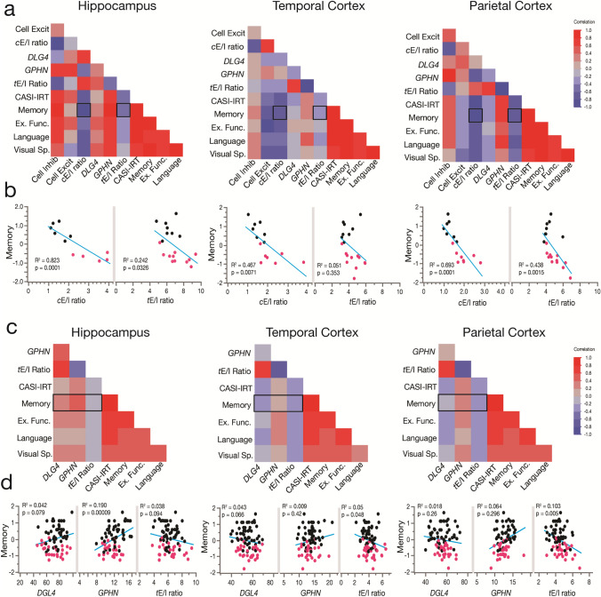
The cellular and synaptic E/I balance in the hippocampus, TCx, and PCx correlates with cognitive performance. a, b Weighted multivariate analysis showing correlation maps for cognitive assessments of CTRL (n = 8) and AD (n = 12) individuals with cellular and synaptic excitatory and inhibitory markers and their ratio from the hippocampus, TCx and PCx. Individuals were scored with the Cognitive Abilities Screening Instrument calibrated using item response theory (CASI-IRT), memory, executive function (Ex. Func), language and visuospatial performance (Visual Sp.). See the cognitive scores section for more information about the use of weight in the analyses. The hippocampus, TCx and PCx showed cellular (cE/I) and transcriptional synaptic E/I (tE/I) ratios correlated with the memory performance of the individuals (p values from Pearson’s correlations). c, d Pearson’s correlation matrix showing cognitive assessments of 86 individuals (56 nondemented, 30 demented AD type) and RNA-seq data of synaptic excitatory (DLG4) and inhibitory (GPHN) markers and their ratio. Hippocampus (n = 75 donors), TCx (n = 79 donors) and PCx (n = 74 donors) (not all the donors had data from the three brain regions). In the hippocampus, memory showed a significant correlation with a reduction in inhibitory synaptic markers and a trend with a loss of excitatory synaptic markers. Cortical regions showed that memory loss better correlates with tE/I ratio increases

