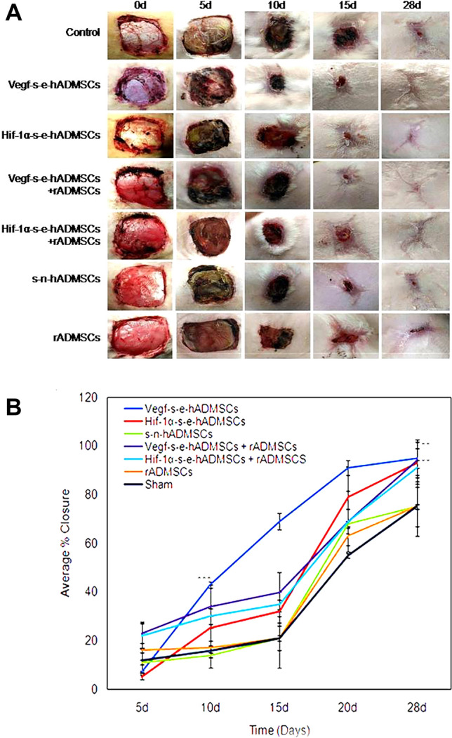
Fig. 4.

A Representative images demonstrating progression of Wound Closure: seven different full thickness excision wound groups in the study of the same dimension (4 cm × 4 cm) were gross imaged on 0 day, 5 days, 10 days, 15 days and upon termination on 28 days. Comparative analysis showed potential of Vegf-s-e-hADMSCs to accelerate the rate of excision wound closure in vivo by 10d. Complete wound closure was observed in s–e-hADMSCs groups, both independently and in combination with rADMSCs at 28 days, (n = 3). B Graphical representation of wounds closure: The wound area measured during the animal evaluation at 0 days, 5 days, 10 days, 15 days and upon termination on 28 days was computed and analyzed. Wound closure demonstrates the effect of seven different types of application highlighting potential of s–e-hADMSCs, both independently and in combination with rADMSCs. Error bars represent standard deviation, ***(P < 0.001); **(P < 0.01); and *(P < 0.05) vs non electroporated hADMSCs, n = 3
