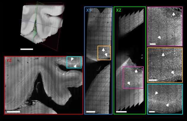
Fig. 3. Higher resolution Mosaic 4 scan of human occipital lobe sample.

3D rendering of a Mosaic 4 acquisitions of a ROI in the vicinity of the human primary visual cortex (left top). Orthogonal views of 50 µm MIPs (XY: blue, XZ: green, YZ: red) shows cortical layers independent of the orientation (white arrows). Jagged edges in XZ view result from deskewing when the edge of the acquisition is inside the tissue. Magnified ROIs of the MIP (XY: yellow, XZ: magenta, YZ: cyan) demonstrate qualitatively the isotropically sampled resolution and image and labelling quality deep within the sample. Scale bars: Volume rendering, XY, and YZ plane 3 mm; XZ: 1.5 mm; magnified ROIs 0.5 mm, respectively.
