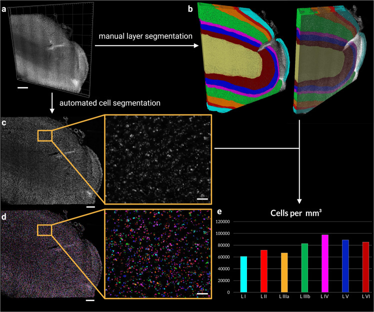
Fig. 5. Segmentation and cell counting in a high resolution (0.725 µm × 0.5127 µm × 0.5127 µm), single-view volume of human brain tissue.
a Volume rendering of the data set used for the cell counting. b Results of the manual layer segmentation for the entire volume. Colours are as follows: Layer I cyan, Layer II bright red, Layer IIIa orange, Layer IIIb green, Layer IV magenta, Layer V blue, Layer VI dark red, and white matter in yellow. The tissue in the unsegmented part of the volume was damaged and did not allow for the segmentation of layers and was excluded from the analysis. Single plane (XY plane of the resliced image volume) of the filtered data (c) and automatically segmented objects (d). Segmented cells are shown in random colours. e Cell density estimates derived from the total count of all the segmented cells per layer segment over the entire image volume (for multiple, unconnected segments of the same layers, the counts were pooled). Colours as indicated for (b). The scale bars for all the images in the left-most side are 500 µm and for the two zoomed-in panels 75 µm.

