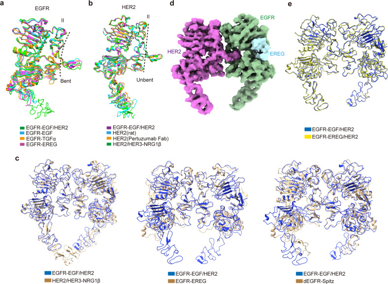
Fig. 2. Structural comparison of different HER family proteins.
a Superposition of the individual EGFR subunit structures in different dimers. EGFR in our EGFR/HER2 dimer (green) is superimposed with the bent EGFR protomer structures in EGF (cyan; PDB code: 1IVO)-, TGFα (orange; PDB code: 1MOX)-, and EREG (magenta; PDB code: 5WB7)-bound EGFR dimers. The RMSDs between our EGFR and the other three structures are 1.82 Å, 2.20 Å, and 1.72 Å, respectively. b Superposition of the individual HER2 subunit structures. HER2 in our EGFR/HER2 dimer (magenta) is overlaid with its monomer structure (rat) (cyan; PDB code: 1N8Y), as well as its structure in complex with Pertuzumab Fab (orange; PDB code: 1S78) or HER3 (green; PDB code: 7MN5). The RMSDs between our HER2 and the other three structures are 1.41 Å, 2.15 Å, and 1.34 Å, respectively. c Superposition of our EGFR/HER2 structure (blue) with the currently reported three asymmetric HER dimers (golden), namely, NRG1β-bound HER2/HER3 (left; PDB code: 7MN5; RMSD 1.95 Å), EREG-bound EGFR (middle; PDB code: 5WB7; RMSD 1.73 Å), and Spitz-bound Drosophila EGFR (dEGFR) (right; PDB code: 3LTG; RMSD 2.68 Å). d Cryo-EM map of the EREG-bound EGFR/HER2 ectodomain complex. EGFR, EREG, and HER2 are colored green, light blue, and magenta, respectively. e Superposition of the EGF (blue)- and EREG (yellow)-bound EGFR/HER2 structures.

