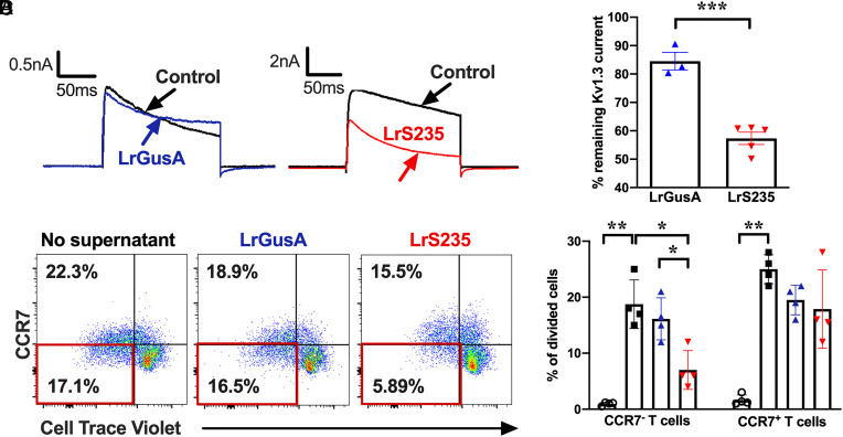
Fig. 1.
Supernatants from LrS235, but not from LrGusA, block Kv1.3 currents and inhibit the proliferation of human CCR7− TEM cells. (A) Representativewhole-cell recordings of L929 cells stably expressing mKv1.3 before (control) and after the addition of supernatants diluted 1/10 of LrGusA or LrS235. (B) Percentage of remaining mKv1.3 currents after addition of LrGusA ( ) or LrS235 (
 ) supernatants diluted 1/10. Mean ± SEM, each data point represents a different measurement. (C) Representative flow cytometry plots of CellTrace Violet dye dilution and CCR7 expression of CD3+ cells from human peripheral blood MNC stimulated for 7 d without any bacterial supernatants (Left) or in the presence of supernatants from LrGusA (Middle) or LrS235 (Right). (D) Percent of divided human CCR7− TEM and CCR7+ naïve/TCM cells in the absence of stimulation (
 ) and after anti-CD3 induced stimulation in the presence of Lr medium (■) or supernatants of LrGusA (
 ) or LrS235 (
 ) diluted 1/100. Mean ± SEM, N = 4 different buffy coat donors. *P < 0.05, **P < 0.01.

