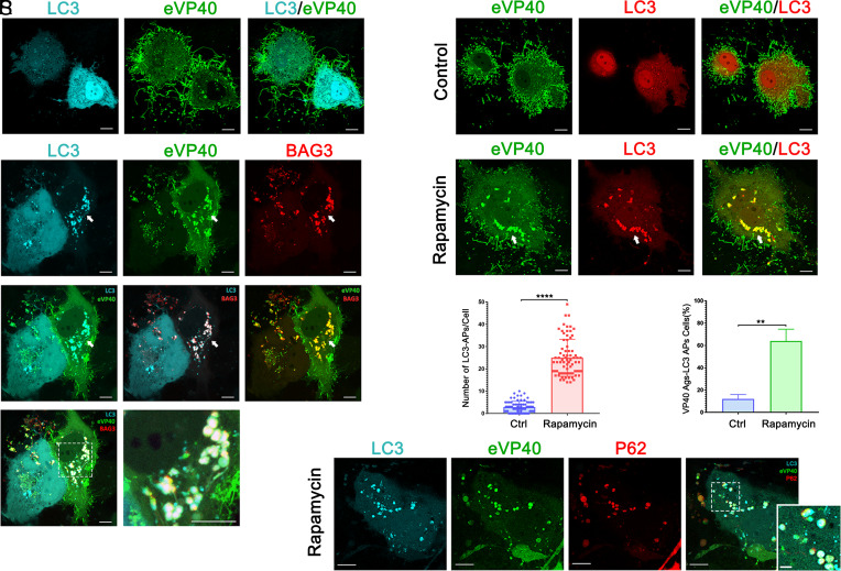
Fig. 4.
Confocal Microscopy showing that BAG3 sequesters and delivers VP40 into LC3-containing autophagosomes. (A–K) Confocal images of Huh7 cells expressing the indicated combinations of proteins including LC3-CFP (cyan), YFP-eVP40 (green), BAG3-mCherry (red). Panel J and zoomed-in panel K highlight the colocalization of LC3, BAG3, and eVP40 in aggregates and autophagosomes. (L–Q) Representative confocal images of Huh7 cells co-expressing LC3-mCherry (red) and GFP-eVP40 (green) in the absence or presence of rapamycin (200 nM). Graphs R and S, Quantification of LC3-containing autophagosomes (APs) measured by the numbers of LC3 puncta per cell (R), and the percentage of cells showing colocalization of VP40 aggregates (Ags) and LC3 APs (S). n ≥ 75 cells were scored for each condition from three independent experiments. Statistical significance was determined by Welch’s t test, ****P < 0.0001, **P < 0.01. (T–W) Confocal images of rapamycin (200 nM)-treated Huh7 cells co-expressing LC3-CFP (cyan), YFP-eVP40 (green) and p62-mCherry (red). (Scale bar, 10 µm in main panels and 2 µm in Inset panels.)

