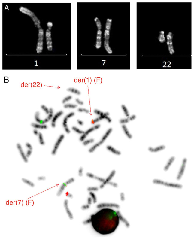
Figure 3.

(A) Partial R-banded karyotype illustrating the translocation t(1;7;22)(p13;q21;q13). The normal chromosomes are shown on the left-hand side and the abnormal chromosomes are on the right-hand side. (B) Metaphase fluorescence in situ hybridization using the RBM15-MKL1 dual fusion translocation probe. Fusion signals were observed on the derivative chromosomes 1 and 7. A normal RBM15 (green) and MKL1 (red) signal were observed on chromosome 1 and 22, respectively. Der, derivative.
