. 2023 Feb 14;13(9):5609–5618. doi: 10.1039/d2ra06859a

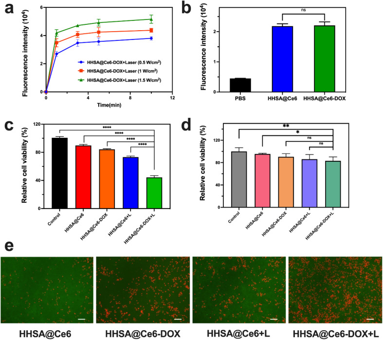
Fig. 5. (a) The fluorescence intensity changes of SOSG with different times between different groups (Ce6 was set to 1 μg mL−1). (b) The fluorescence intensity of DCFH-DA in the HHSA@Ce6 and HHSA@Ce6-DOX groups (0.625 μg mL−1 Ce6 equiv.) in 4T1 cells. Relative cell viability of 4T1 cells (c) and 293T (d) cells before and after the irradiation (+L) in different treatment groups (0.625 μg mL−1 Ce6 equiv.). (e) Fluorescent images of live (green) and dead (red) cells before and after the irradiation (+L) in different treatment groups (0.625 μg mL−1 Ce6 equiv.) in 4T1 cells. Scale bar: 100 μm. P values: ns P > 0.05, *P < 0.05, **P < 0.01, ***P < 0.001, ****P < 0.0001 were calculated by SPSS with a t-test.

Fig. 5