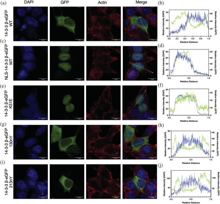
FIGURE 7.

Subcellular localization of nitrated 14‐3‐3. HEK293T cells expressing 14‐3‐3 β‐eGFP fusion proteins (green channel) were stained for the nucleus with DAPI (blue channel) and for actin with Alexa Flour 647 Phalloidin (red channel) and imaged by confocal microscopy. Subcellular fluorescence profile quantification of DAPI and eGFP are shown in far‐right panels. (a, b) 14‐3‐3 β‐eGFP WT, (c, d) NLS‐14‐3‐3 β‐eGFP WT, (e, f) 14‐3‐3 β‐eGFP K51E, (g, h) 14‐3‐3 β‐eGFP 130nY, and (i, j) 14‐3‐3 β‐eGFP 213nY. See Figure S9 for uncropped images.
