. 2023 Feb 7;307(2):e222557. doi: 10.1148/radiol.222557

Figure 2:

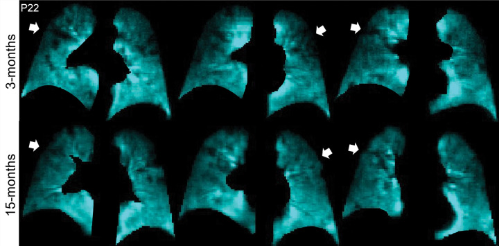
129Xe MRI ventilation scans 3 months (top) and 15 months (bottom) after COVID-19 infection in a 65-year-old man who was not hospitalized during acute infection. Coronal 129Xe MRI scans show lung sections (cyan), with arrows indicating MRI ventilation abnormalities that improved at the 15-month follow-up. At 3 and 15 months, the forced expiratory volume in 1st second of expiration percent predicted was 95% and 102%, diffusing capacity of lung for carbon monoxide percent predicted was 97% and 105%, St George Respiratory Questionnaire score was 36 and 27, and ventilation defect percent was 7% and 2%, respectively.

129Xe MRI ventilation scans 3 months (top) and 15 months (bottom) after COVID-19 infection in a 65-year-old man who was not hospitalized during acute infection. Coronal 129Xe MRI scans show lung sections (cyan), with arrows indicating MRI ventilation abnormalities that improved at the 15-month follow-up. At 3 and 15 months, the forced expiratory volume in 1st second of expiration percent predicted was 95% and 102%, diffusing capacity of lung for carbon monoxide percent predicted was 97% and 105%, St George Respiratory Questionnaire score was 36 and 27, and ventilation defect percent was 7% and 2%, respectively.