. 2023 Feb 7;307(2):e222557. doi: 10.1148/radiol.222557

Figure 5:

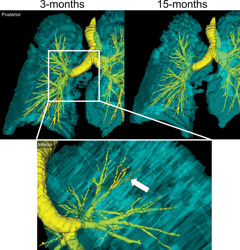
Representative images show a three-dimensional model of 129Xe MRI lung sections (cyan) and CT airways (yellow) in a 65-year-old man who was not hospitalized during acute COVID-19 infection. Top: Posterior views show ventilation abnormalities at 3 months (left), which resolved at 15 months (right). Bottom: Inset shows the inferior view, with the arrow pointing to an airway LB3 ventilation defect at 3 months.

Representative images show a three-dimensional model of 129Xe MRI lung sections (cyan) and CT airways (yellow) in a 65-year-old man who was not hospitalized during acute COVID-19 infection. Top: Posterior views show ventilation abnormalities at 3 months (left), which resolved at 15 months (right). Bottom: Inset shows the inferior view, with the arrow pointing to an airway LB3 ventilation defect at 3 months.