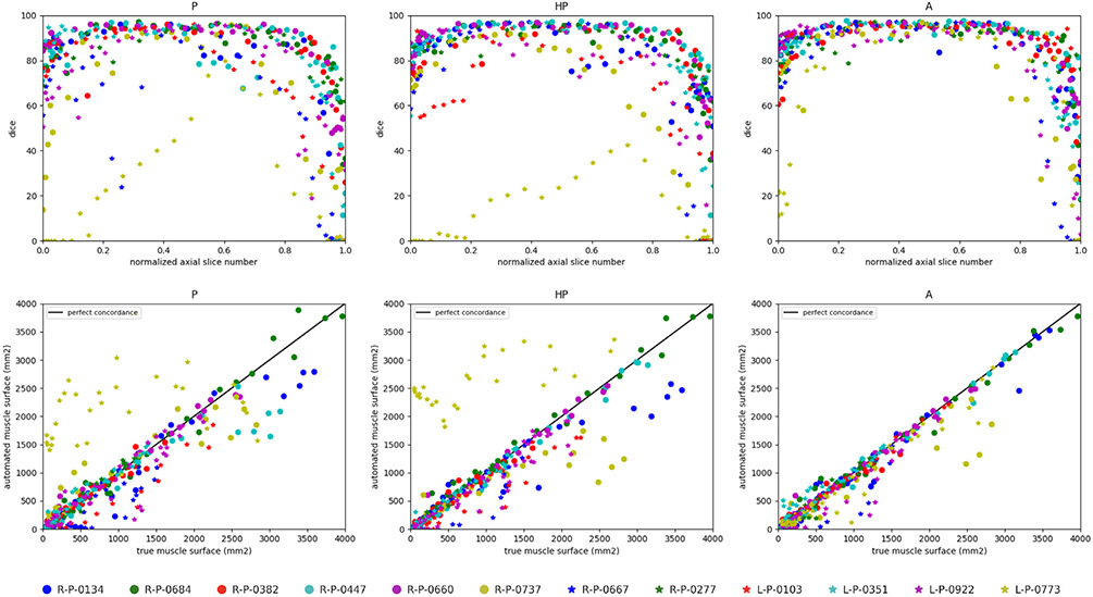
Fig. 5.
Deltoid segmentation accuracy using U-Net (Ronneberger et al., 2015) with learning schemes P, HP and A for each annotated slice of the whole pathological dataset. Top raw shows Dice scores (%) with respect to the normalized axial slice number obtained by linearly scaling slice number from [zmin, zmax] to [0, 1] where {zmin, zmax} are the minimal and maximal axial slice indices displaying the deltoid. Bottom row displays concordance between groundtruth and predicted deltoid muscle surfaces in mm2. Black line indicates perfect concordance.

