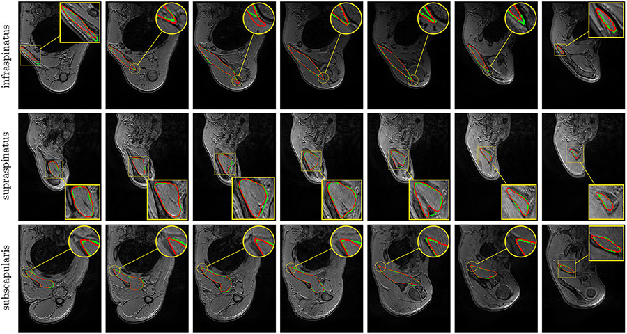
Fig. 7.
Automatic pathological segmentation of infraspinatus, supraspinatus and subscapularis using U-Net (Ronneberger et al., 2015) with training on both healthy and pathological data simultaneously (A). Groundtruth and estimated delineations are in green and red respectively. Displayed results cover the whole muscle spatial extents for R-P-0447 (top), R-P-0660 (middle) and R-P-0134 (bottom) examinations.

