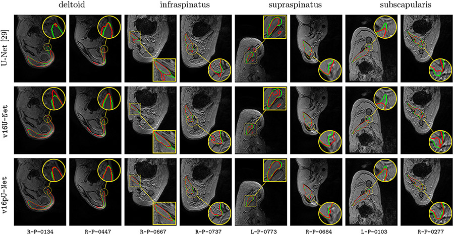
Fig. 9.
Automatic pathological segmentation of deltoid, infraspinatus, supraspinatus and subscapularis using U-Net (Ronneberger et al., 2015), v16U-Net and v16pU-Net with training on both healthy and pathological data simultaneously (A). Groundtruth and estimated delineations are in green and red respectively. 8 pathological examinations among the 12 available are involved to provide valuable insight into the overall performance.

