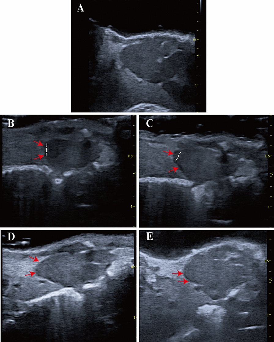
Fig. 3.

Echocardiographic imaging can monitor regeneration of X. tropicalis injured hearts in a scar-free manner. A: Representative echocardiography image of the same-age nonapical resection group under B-mode. B: Representative image 5 days after apical resection. The damaged heart with a missing apex (left side of the dashed line) was clearly identified under echocardiography. C: Representative image 10 days after apical resection. D: Representative image 30 days after apical resection. E: Representative image 45 days after apical resection. The regeneration of the injured heart was able to be monitored and justified dynamically by the recovery of morphology and anatomic structure under echocardiographic imaging at 5 days, 10 days, 30 days and 45 days after apical resection. The boundary between the apical region of the regenerated heart and the surrounding tissue was clear, and no adhesion with the surrounding tissue was found at 30 days and 45 days after apical resection. Red arrow: Area of the boundary between the apical region of the regenerated heart and the surrounding tissue. Dashed line: Boundary of the regeneration zone and noninjury zone
