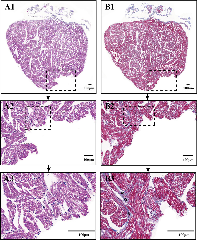
Fig. 7.
Histology analysis confirms the accuracy of echocardiography assessment for nonperfect regeneration with the adhesion of injured X. tropicalis hearts. A1: Representative H&E staining of regenerated hearts at 45 daar; echocardiography assessment identified regeneration in a nonperfect manner with adhesion. A2: 5 × of dotted rectangle area of A1. A3: 2.4 × of dotted rectangle area of A2.. B1: Representative Masson’s trichrome staining of a regenerated heart at 45 daar; echocardiography assessment identified regeneration in a nonperfect manner with adhesion. B2: 5 × of dotted rectangle area of B1. B3: 2.4 × of dotted rectangle area of B2. H&E staining showed that the amputated heart was regenerated with a nearly heart-shaped morphology containing a defect after the adhesion was cleaned. In addition, Masson’s trichrome staining revealed fibrotic structures in the regenerated myocardium. As the adhesion between the regenerated area and peripheral tissue was cleaned when the regenerated hearts were isolated, the fibrotic tissue of the adhesion could not be stained

