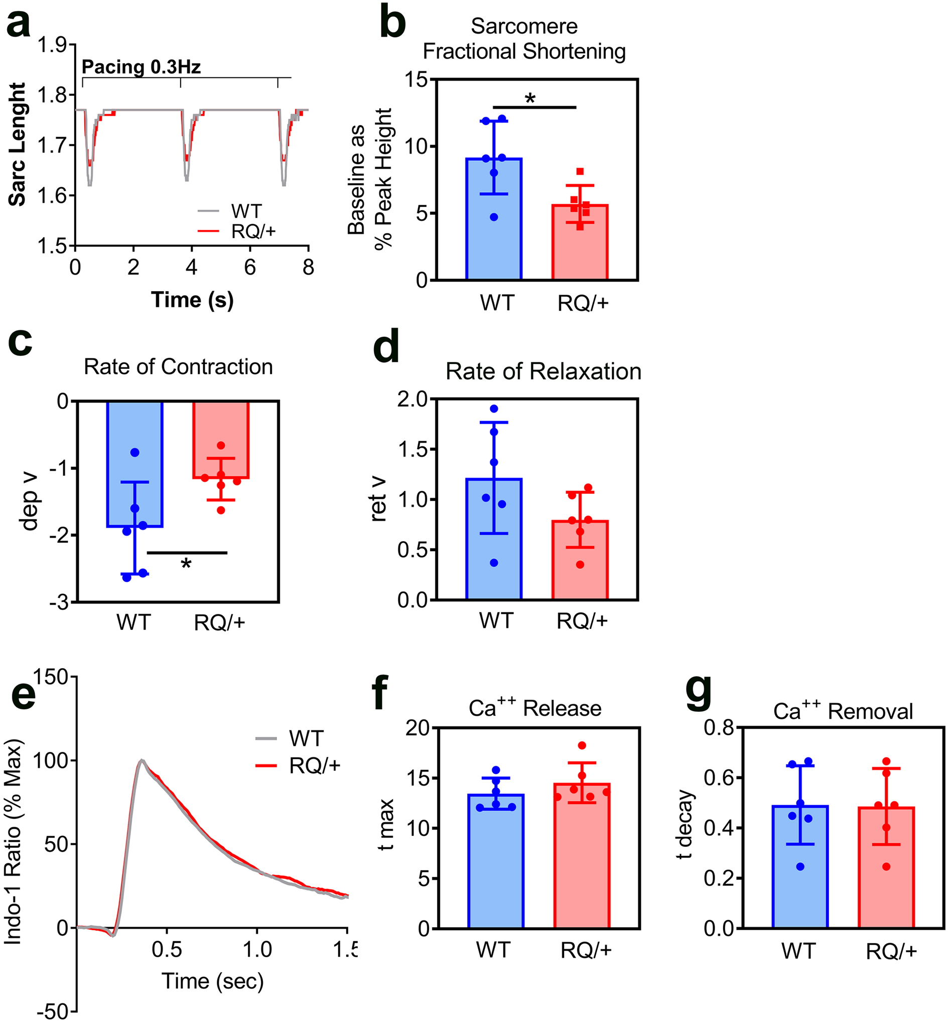
Fig. 2: Contractility and cytosolic Ca++ transients in adult cardiac myocytes isolated from Prkg1RQ/+ mice and control litter mates.

(a-d) Cardiac myocyte contractility was assessed by monitoring sarcomere length upon electrical stimulation (20V, 0.3 Hz). Representative traces (a) from three consecutive contractions are shown for wild type (WT, grey) and Prkg1RQ/+ (RQ/+, red) myocytes. Sarcomere fractional shortening (b) was measued as a percent of sarcomere length change during contraction (baseline as a percent of peak height). Rate of contraction (c) is expressed as departure velocity (dep v). Rate of relaxation (d) is expressed as return velocity (ret v), i.e. velocity of the return phase of the transient. (e-g) Representative cytosolic Ca++ transients (e) were recorded in paced-contracting myocytes from wild type (WT, grey) and Prkg1RQ/+ (RQ/+, red) mice; cells were pre-loaded with Indo-1 for 30 min. Rates of Ca++ release into the cytosol (f, t max) and rates of Ca++ removal from the cytosol (g, t decay) were measured. The data in panels b-d and f-g represent n=6 mice per genotype with ≥20 cells analyzed per mouse; means +/− SD with *p<0.05 for the indicated comparisons by two-sided t-test.
