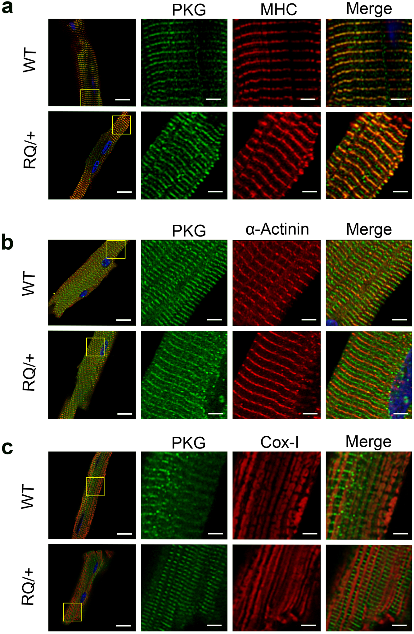
Fig. 3: Subcellular localization of wild type and mutant PKG1RQ in cardiac myocytes.

Cardiac myocytes were isolated from adult (6 mo old) wild type (WT) and Prkg1RQ/+ (RQ/+) mice, and were allowed to adhere to laminin-coated glass coverslips. The cells were fixed and stained, and proteins were visualized by immunofluorescence. Green staining represents PKG1; cells were co-stained (red) for either myosin heavy chain (MHC) representing the M-band (a), α-actinin representing the Z-band (b), or cytochrome oxidase-1 (Cox-1) highlighting mitochondria (c). Nuclei were counterstained with Hoechst 33342 (blue). Images were acquired on a Zeiss LSM880 fluorescent confocal microscope with Airyscan using a 60x objective, and are representative for cardiomyocyte preparations from n=3 mice per genotype, with at least 5–10 cells analyzed per mouse. (bars in left column represent 20 μm, in magnified views 4 μm).
