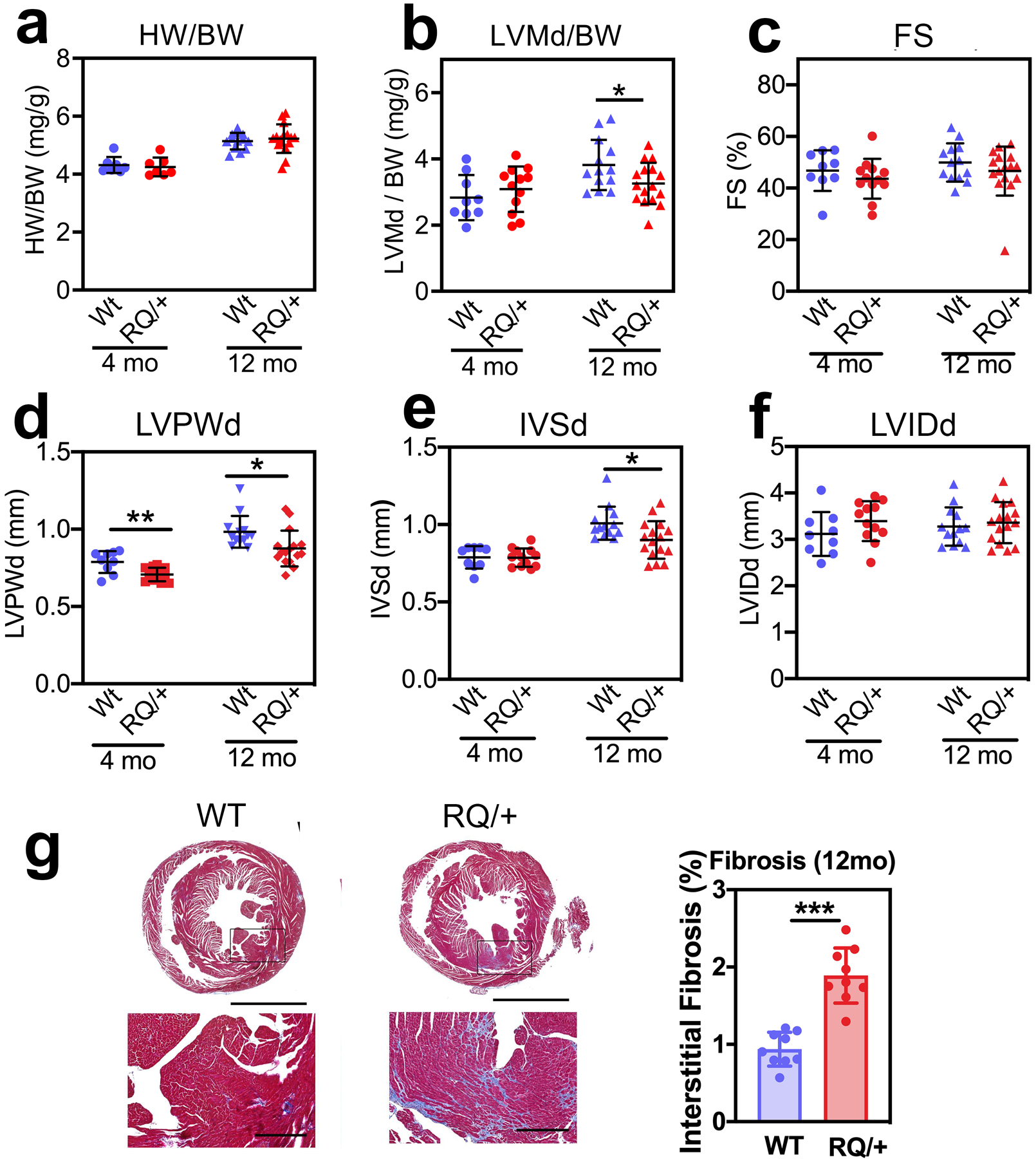
Fig. 4: Basal cardiac phenotype of aging Prkg1RQ/+ mice and control litter mates.

(a,b) Heart weight (HW) and left ventricular mass in diastole (LVMd) were normalized to body weight (BW) in 4-month and 12-month-old male Prkg1RQ/+ (RQ/+) mice and wild type (WT) litter mates. (c-f) Cardiac parameters were measured by echocardiography in 4-month and 12-month-old male WT and RQ/+ mice: (c) LV fractional shortening (FS); (d) LV posterior wall thickness in diastole (LVPWd); (e) inter-ventricular septum thickness in diastole (IVSd); (f) LV inner diameter in diastole (LVIDd). (g) Cross-sections of the hearts from 12-month-old male mice stained with trichrome to highlight collagen in blue. Interstitial fibrosis was quantified using Image-Pro Premier software and is expressed as percent of cross-sectional area. Data are shown as mean ± SD for n=9 and 13 WT mice and n=12 and 16 RQ/+ mice in the 4- and 12-month-old groups, respectively, except for panel a with seven 4-month-old mice and panel g with nine 12-month-old mice per genotype. *p<0.05 for the indicated comparisons by 2-Way ANOVA (panels a-f), ***p<0.001 by two-sided t-test (panel g).
