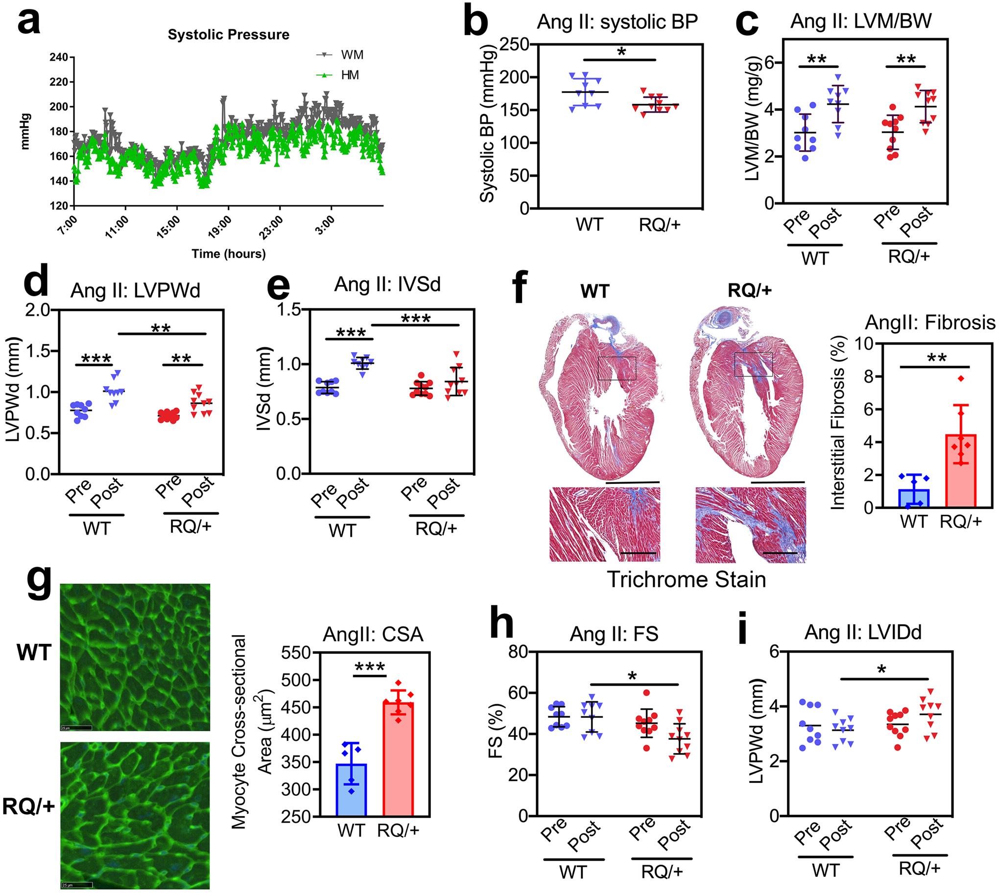
Fig. 5: Cardiac remodeling and function in response to angiotensin II.

Four month-old male Prkg1RQ/+ (RQ/+) mice and wild type (WT) litter mates received Ang II at 1 μg/kg/min subcutaneously per osmotic minipump for three weeks. (a) Systolic blood pressure was measured by telemetry over 24 h, starting after 2 weeks of Ang II infusion (n=4 mice per genotype). (b) Systolic blood pressure was measured by tail cuff plethysmography after 2 weeks of Ang II infusion in mice that underwent echocardiography after 3 weeks (n=9 WT and n=10 RQ/+). (c-e, h-i) Cardiac parameters were measured by echocardiography after three weeks of Ang II infusion (n=9 WT and n=10 RQ/+): (c) LV mass (LVM) normalized to body weight (BW); (d) LV posterior wall thickness in diastole (LVPWd); (e) inter-ventricular septum thickness in diastole (IVSd); (h) LV fractional shortening (FS); and (i) LV inner diameter in diastole (LVIDd). (f) Collagen content was quantified on trichrome-stained sections as in Fig. 4g (n=5 WT and n=7 RQ/+ mice). (g) Cardiac myocyte cross-sectional area (CSA) was measured on wheat germ agglutinin-stained cross sections (n=5 WT and n=7 RQ/+ mice). Graphs represent means ± SD; *p<0.05, **p<0.01, and ***p<0.001 for the indicated comparisons by 2-Way ANOVA (panels c-e, h,i), or by two-sided t-test (panels b, f, g).
