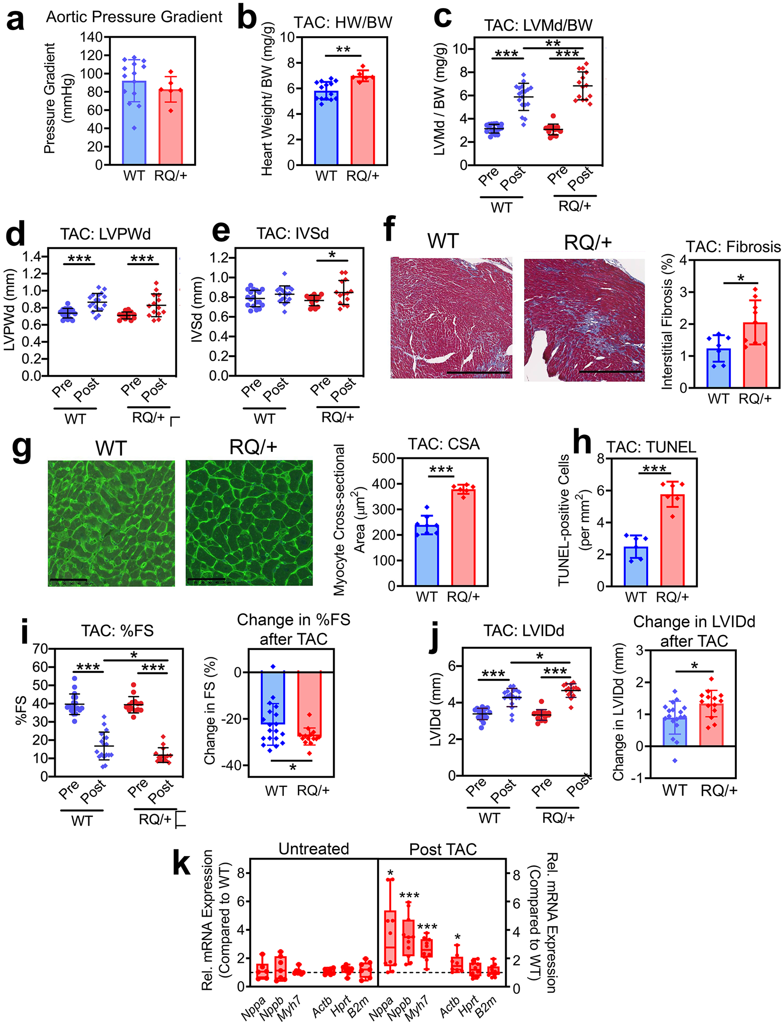
Fig. 6: Cardiac remodeling and function in response to transaortic constriction (TAC).

Four to five month-old male Prkg1RQ/+ (RQ/+) mice and wild type (WT) litter mates were subjected to transaortic constriction and were euthanized 2 weeks later. (a) The aortic pressure gradient generated by TAC was measured proximal and distal to the constriction by invasive blood pressure monitoring at the time of euthanasia. (b) Heart weight (HW) was normalized to body weight (BW). (c-e, i,j) Cardiac echocardiography was performed two days before (Pre) and 14 d after TAC (Post) in n=18 WT and n=14 RQ/+ mice: (c) LV mass in diastole (LVMd) normalized to body weight (BW); (d) left ventricular posterior wall thickness in diastole (LVPWd); (e) inter-ventricular septum thickness in diastole (IVSd);); (i) left ventricular fractional shortening (FS); and (j) left ventricular inner diameter in diastole (LVIDd). (f) Collagen content was quantified on trichrome-stained sections as in Fig. 4g (n=7 mice per genotype). (g) Cardiac myocyte cross-sectional area (CSA) was measured on wheat germ agglutinin-stained cross sections (n=7 mice per genotype). (h) Cardiac myocyte apoptosis was assessed by TUNEL staining (n=6 mice per genotype). (k) Expression of hypertrophic genes and house-keeping genes in hearts from RQ/+ mice was compared to WT mice under basal conditions (untreated) and after TAC. Gene expression was measured by quantitative RT-PCR, and was normalized to 18S RNA. For each gene, mean ΔCT values in the wild type group were assigned a value of one (n= 10 mice per genotype). Data in a-j are plotted as means ± SD; *p<0.05, **p<0.01, and ***p<0.001 for the indicated comparisons by two-tailed t-test (b, f-h), one-spample t-test (k), or 2-Way ANOVA (c-e, i,j).
