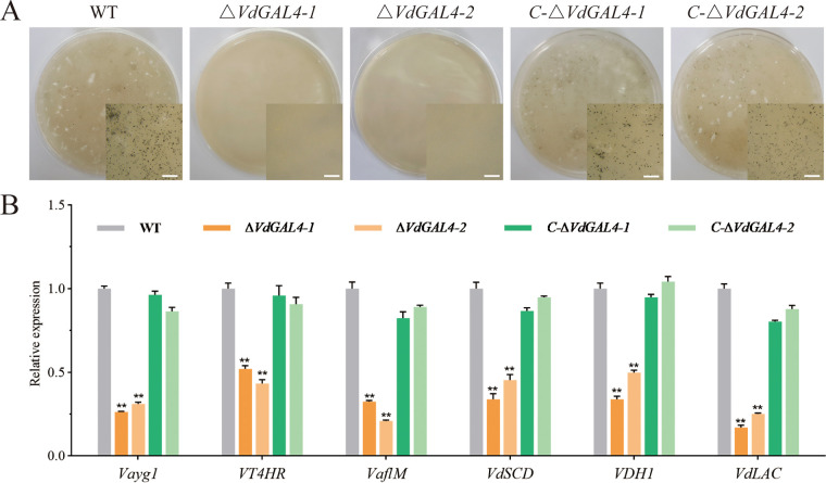
FIG 5.
Assays of the role of VdGAL4 in the formation of microsclerotium and melanin. (A) Microsclerotium formation of WT, ΔVdGAL4, and C-ΔVdGAL4 strains cultured in BMM medium for 40 days. The microsclerotium in the right bottom corner is shown by microscopy. Bar, 1 mm. (B) Assays of the relative expression of melanin-related genes by RT-qPCR. The error bar represents standard error of the mean. **, P < 0.01.

