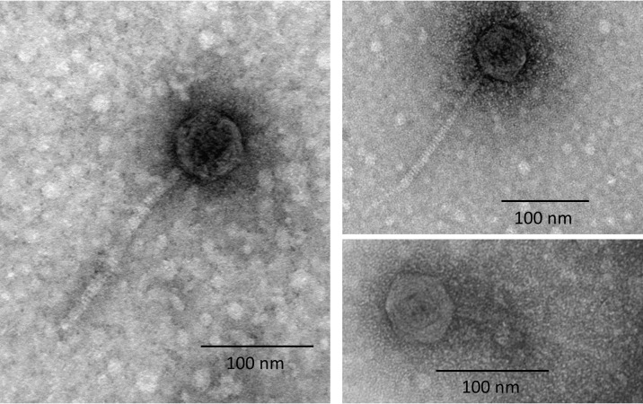
FIG 2.
Transmission electron microscopy of the Φ1207.3 phage particles. TEM observation after negative staining of phage preparations shows the presence of phage particles with a morphology consistent with a siphovirus. The capsid shows an icosahedral symmetry and has a diameter of 62 nm, while the tail is noncontractile and 175 nm (±1 nm) long. The scale is reported for each panel.

