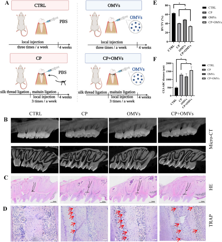
FIG 2.
P. gingivalis OMVs promote alveolar bone resorption in vivo. (A) Schematic diagram of animal experiments. (B) The level of the rat alveolar bone resorption of four groups was measured by micro-CT and (C) H&E staining. The distances from the CEJ to ABC are marked in black. (D) TRAP staining. Red arrows indicate TRAP-positive surfaces. (E and F) The percentage of bone volume over tissue volume (BV/TV) and the distance from the cementoenamel junction (CEJ) to the alveolar bone crest (ABC) were calculated. Data are shown as mean ± SD with n = 5 or 6 rats per group. Data between two groups were compared using Student's t test. *, P < 0.05.

